. 2021 Nov 22:12:762121.
doi: 10.3389/fpls.2021.762121. eCollection 2021.

Mapping Pectic-Polysaccharide Epitopes in Cell Walls of Forage Chicory (Cichorium intybus) Leaves

Affiliations

Mapping Pectic-Polysaccharide Epitopes in Cell Walls of Forage Chicory (Cichorium intybus) Leaves

Xuezhao Sun et al. Front Plant Sci. .

Abstract

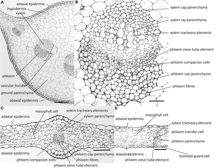
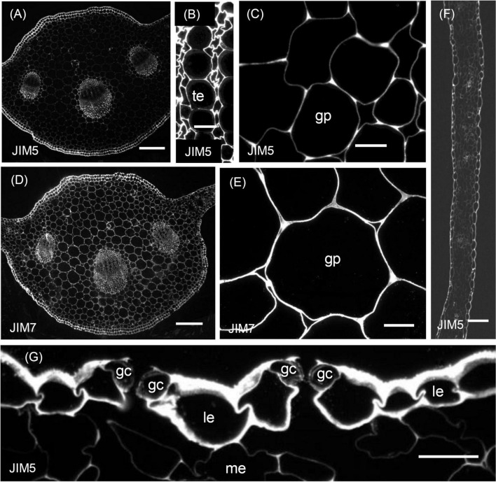
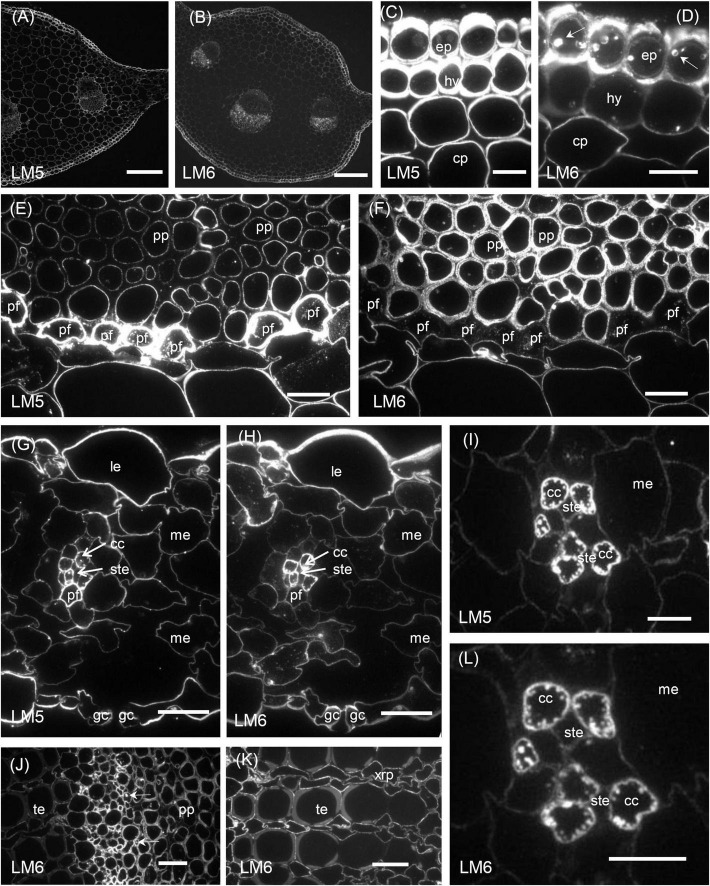
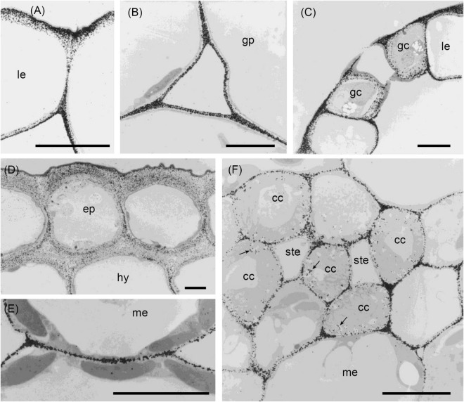
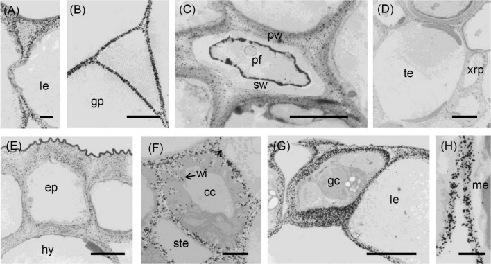
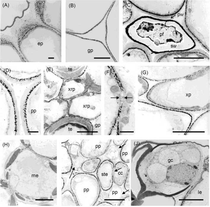
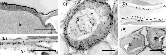
The cell walls of forage chicory (Cichorium intybus) leaves are known to contain high proportions of pectic polysaccharides. However, little is known about the distribution of pectic polysaacharides among walls of different cell types/tissues and within walls. In this study, immunolabelling with four monoclonal antibodies was used to map the distribution of pectic polysaccharides in the cell walls of the laminae and midribs of these leaves. The antibodies JIM5 and JIM7 are specific for partially methyl-esterified homogalacturonans; LM5 and LM6 are specific for (1→4)-β-galactan and (1→5)-α-arabinan side chains, respectively, of rhamnogalacturonan I. All four antibodies labelled the walls of the epidermal cells with different intensities. JIM5 and JIM7, but not LM5 or LM6, labelled the middle lamella, tricellular junctions, and the corners of intercellular spaces of ground, xylem and phloem parenchyma. LM5, but not LM6, strongly labelled the walls of the few sclerenchyma fibres in the phloem of the midrib and lamina vascular bundles. The LM5 epitope was absent from some phloem parenchyma cells. LM6, but not LM5, strongly labelled the walls of the stomatal guard cells. The differential distribution of pectic epitopes among walls of different cell types and within walls may reflect the deposition and modification of these polysaccharides which are involved in cell wall properties and cell development.

Keywords: arabinan; chicory; galactan; homogalacturonan; immunolabelling; pectins.


Conflict of interest statement

The authors declare that the research was conducted in the absence of any commercial or financial relationships that could be construed as a potential conflict of interest.

Figures

FIGURE 1
FIGURE 1
Photomicrographs of transverse sections cut from a rosette leaf of an 8-week-old chicory plant. (A) A transverse section of the midrib. (B) A vascular bundle in the midrib. (C) A transverse section of the lamina with a major vein. (D) A transverse section of the lamina with a minor vein. Although it is difficult to identify the transfer cells, sieve tube elements, and companion cells in the photomicrographs, the lines point to where these cell types occur. Bars: (A) 200 μm; (B) 40 μm; and (C,D) 100 μm.
FIGURE 2
FIGURE 2
Immunofluorescence micrographs of transverse sections cut from a rosette leaf from an 8-week-old chicory plant and labelled with JIM5 or JIM7. (A) The midrib labelled with JIM5. (B) Xylem tracheary elements and adjacent xylem parenchyma cells in a vascular bundle in the midrib labelled with JIM5. (C) Parenchyma cells (ground parenchyma) between the midrib hypodermis and the vascular bundles, showing more intense JIM5 labelling at the cell corners. (D) The midrib labelled with JIM7. (E) Parenchyma cells (ground parenchyma cells) between the midrib hypodermis and the vascular bundles labelled with JIM7. (F) The lamina, showing moderate labelling of the outer epidermal walls and the walls of cells in the minor veins. (G) Stomatal guard cells, showing weak labelling of the walls of stomatal guard cells, with the outermost regions of these walls and outer ledge more intensely labelled. gp: ground parenchyma; gc: stomatal guard cells; le: lamina epidermis; me: mesophyll; and te: tracheary element. Bars: (A,D) 200 μm; (B,C,E,G) 100 μm.
FIGURE 3
FIGURE 3
Immunofluorescence micrographs of transverse sections cut from a rosette leaf of an 8-week-old chicory plant and labelled with LM5 or LM6. (A) The midrib labelled withLM5. (B) The midrib labelled with LM6. (C) The midrib labelled with LM5, showing very strong labelling on the epidermal and hypodermal walls and moderate labelling of the parenchyma cell walls. (D) The midrib labelled with LM6, showing the very strong labelling of the epidermal walls and the moderate labelling of the hypodermal and parenchymal cell walls. (E) The midrib labelled with LM5, showing very strong labelling of the phloem fibre walls and the inner walls of the phloem parenchyma cells. (F) The midrib labelled with LM6, showing strong labelling of the phloem parenchyma cell walls but not the middle lamellae between these cells and no labelling of the phloem fibre walls. (G) The lamina labelled with LM5, showing strong labelling of the epidermal cell walls and no labelling on the walls of the stomatal guard cells. (H) The lamina labelled with LM6, showing intense labelling of the outer walls of the epidermal cells and the walls of the phloem transfer cells in a minor vein. (I) A minor vein in the lamina labelled with LM5, showing strong labelling of the walls and wall ingrowths of phloem companion cells (transfer cells). (J) The phloem in a midrib vascular bundle labelled with LM6, showing labelling of inclusions in some inner phloem cells. (K) The xylem in a midrib vascular bundle labelled with LM6, showing no labelling of the secondary walls of the tracheary elements but labelling of the primary walls of adjacent elements, and the walls of xylem ray parenchyma cells. (L) A minor vein in a lamina labelled with LM6, showing strong labelling of the walls and wall ingrowths of phloem transfer cells. cc: phloem transfer cell; ep: epidermal cell; gc: stomatal guard cell; hy: hypodermis; le: lamina epidermis; me: mesophyll; pf: phloem fibre; pp: phloem parenchyma; ste: sieve tube element; te: tracheary element; and xrp: xylem ray parenchyma. Bars: (A,B) 200 μm; (C–H,J,K) 20 μm; and (I,L) 5 μm.
FIGURE 4
FIGURE 4
Transmission electron micrographs of transverse sections cut from a rosette leaf of an 8-week-old chicory plant and immunogold labelled with JIM5. (A) A lamina epidermal cell, showing labelling in the outer wall increasing toward the outer surface. (B) The ground parenchyma in the midrib, showing labelling particularly concentrated at the cell corners and along walls near the middle lamella. (C) Stomatal guard cells, showing strong labelling of the outermost regions of the cell walls and throughout the outer ledge and no labelling of the cuticle. (D) Epidermis and hypodermis, showing heavy labelling of the middle region of walls between two epidermal cells and between two hypodermal cells. (E) Mesophyll cells, showing labelling of the walls that is particularly strong in regions where adjacent cells adhere. (F) A minor vein in a lamina, showing labelling of the wall ingrowths of transfer cells. cc: transfer cell; ep: epidermal cell; gc: stomatal guard cell; gp: ground parenchyma; hy: hypodermis; le: lamina epidermis; me: mesophyll; ste: phloem sieve tube element; and →: wall ingrowth. Bars: 5 μm.
FIGURE 5
FIGURE 5
Transmission electron micrographs of transverse sections cut from a rosette leaf of an 8-week-old chicory plant and immunogold labelled with JIM7. (A) A lamina epidermal cell, showing the walls evenly labelled. (B) A ground parenchyma cell in the midrib, showing labelling throughout the walls but less intense at the corners of cell junctions. (C) A phloem fibre, showing labelling in the primary walls but no labelling in the secondary walls. (D) The xylem in the midrib, showing labelling in the primary walls of the tracheary elements and xylem ray parenchyma cells but no labelling in the secondary walls of the tracheary elements. (E) Epidermal and hypodermal cells, showing even labelling of the walls. (F) Transfer cells in a minor vein of the lamina, showing labelling of transfer cell walls and wall ingrowths. (G) Stomatal guard cells, showing dense, but non-uniform labelling of the guard cell walls, with labelling also present in the cuticular layer. (H) Mesophyll cells, showing no labelling at the corners of intercellular spaces. cc: transfer cell; gp: ground parenchyma; ep: epidermal cell; gc: stomatal guard cell; hy: hypodermis; le: lamina epidermis; me: mesophyll; pf: phloem fibre; pw: primary wall; sw: secondary wall; te: tracheary element; xrp: xylem ray parenchyma; and wi: wall ingrowth. Bars: (A,F,H) 1 μm; (B–E,G) 5 μm.
FIGURE 6
FIGURE 6
Transmission electron micrographs of transverse sections cut from a rosette leaf of an 8-week-old chicory plant and immunogold labelled with LM5. (A) The outer wall of epidermal cells in the midrib, showing that labelling gradually increased from the cuticle to the plasma membrane, but was largely absent from the middle lamella. (B) A ground parenchyma cell in the midrib, showing labelling of the primary walls, but no labelling of the middle lamella or the corners of the intercellular spaces. (C) A phloem fibre, showing that labelling was heavy and in a narrow region of the primary walls. (D) A parenchyma cell in the phloem cap, showing thick walls with labelling confined to the region near the plasma membrane. (E) The xylem, showing that labelling is absent in the secondary walls of the tracheary elements, but present in the primary walls of tracheary elements and xylem ray parenchyma cells. (F) A ground parenchyma cell, showing no labelling of the pit field. (G) A xylem parenchyma cell, showing the same pattern of labelling as in (B). (H) Mesophyll cells, showing that most walls were labelled, but a few were not. (I) Phloem parenchyma cells near the sieve tube elements, showing no labelling of the walls of some of the phloem parenchyma cells. (J) Stomatal guard cells, showing no labelling of the walls. cc: transfer cell; gp: ground parenchyma; ep: epidermal cell; gc: stomatal guard cell; le: lamina epidermis; me: mesophyll cell; pf: phloem fibre; pp: phloem parenchyma; pw: the primary wall; ste: sieve tube element; sw: the secondary wall; te: tracheary element; xrp: xylem ray parenchyma; and xp: xylem parenchyma; arrows show the absence of labelling. Bars: (A,D,F) 1 μm; (B,C,E,G–J) 5 μm.
FIGURE 7
FIGURE 7
Transmission electron micrographs of transverse sections cut from a rosette leaf of an 8-week-old chicory plant and immunogold labelled with LM6. (A) An epidermal cell in the lamina, showing dense labelling of the outer walls, but light labelling at the junction of two cells. (B) Ground parenchyma cells, showing labelling present in the outer region of the primary walls but no labelling of the corners of intercellular spaces. (C) A phloem parenchyma cell, showing heavy labelling of the walls (D) Mesophyll cells in the lamina, showing labelling in the walls; this labelling appears as two lines in some walls. (E) Stomatal guard cells, showing heavy labelling of the walls. gp: ground parenchyma; gc: stomatal guard cell; le: lamina epidermis; me: mesophyll; and pp: phloem parenchyma. Bars: (A,E): 5 μm; (B–D): 2 μm.

Similar articles

Cited by

References

    1. Andeme-Onzighi C., Girault R., His I., Morvan C., Driouich A. (2000). Immunocytochemical characterization of early-developing flax fiber cell walls. Protoplasma 213 235–245. 10.1007/bf01282161 - DOI
    1. Andersen M. C., Boos I., Marcus S. E., Kračun S. K., Rydahl M. G., Willats W. G., et al. (2016). Characterization of the LM5 pectic galactan epitope with synthetic analogues of β-1,4-D-galactotetraose. Carbohydr. Res. 436 36–40. 10.1016/j.carres.2016.10.012 - DOI - PubMed
    1. Barry T. N. (1998). The feeding value of chicory (Cichorium intybus) for ruminant livestock. J. Agric. Sci. 131 251–257.
    1. Blake A. W., Marcus S. E., Copeland J. E., Blackburn R. S., Knox J. P. (2008). In situ analysis of cell wall polymers associated with phloem fibre cells in stems of hemp, Cannabis sativa L. Planta 228 1–13. 10.1007/s00425-008-0713-5 - DOI - PubMed
    1. Buffetto F., Cornuault V., Rydahl M. G., Ropartz D., Alvarado C., Echasserieau V., et al. (2015). The deconstruction of pectic rhamnogalacturonan I unmasks the occurrence of a novel arabinogalactan oligosaccharide epitope. Plant Cell Physiol. 56 2181–2196. 10.1093/pcp/pcv128 - DOI - PubMed

LinkOut - more resources