

Integration of PEG-conjugated gadolinium complex and superparamagnetic iron oxide nanoparticles as T 1- T 2 dual-mode magnetic resonance imaging probes
- PMID: 34881046
- PMCID: PMC8648151
- DOI: 10.1093/rb/rbab064
Integration of PEG-conjugated gadolinium complex and superparamagnetic iron oxide nanoparticles as T 1- T 2 dual-mode magnetic resonance imaging probes
Abstract
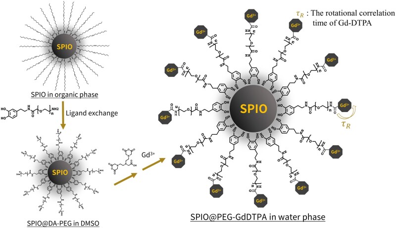
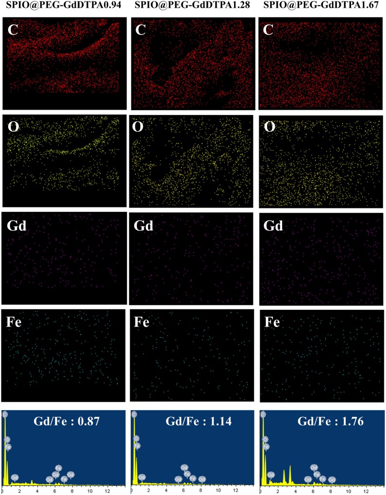
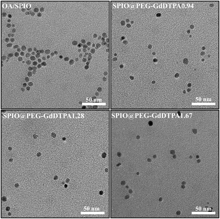
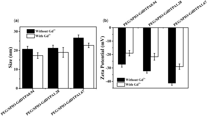
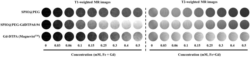
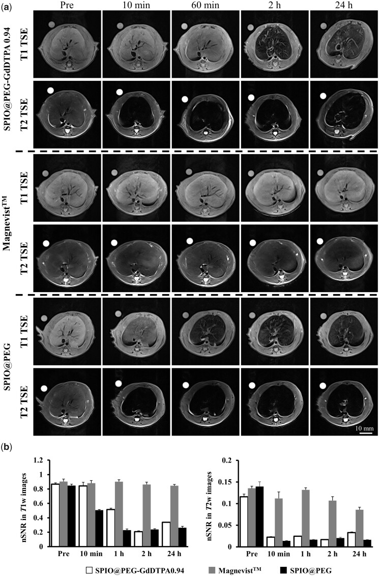
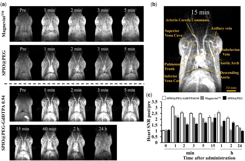
The T 1-T 2 dual-mode probes for magnetic resonance imaging (MRI) can non-invasively acquire comprehensive information of different tissues or generate self-complementary information of the same tissue at the same time, making MRI a more flexible imaging modality for complicated applications. In this work, three Gadolinium-diethylene-triaminepentaaceticacid (Gd-DTPA) complex conjugated superparamagnetic iron oxide (SPIO) nanoparticles with different Gd/Fe molar ratio (0.94, 1.28 and 1.67) were prepared as T 1-T 2 dual-mode MRI probes, named as SPIO@PEG-GdDTPA0.94, SPIO@PEG-GdDTPA1.28 and SPIO@PEG-GdDTPA1.67, respectively. All SPIO@PEG-GdDTPA nanocomposites with 8 nm spherical SPIO nanocrystals showed good Gd3+ chelate stability. SPIO@PEG-GdDTPA0.94 nanocomposites with lowest Gd/Fe molar ratio show no cytotoxicity to Raw 264.7 cells as compared to SPIO@PEG-GdDTPA1.28 and SPIO@PEG-GdDTPA1.67. SPIO@PEG-GdDTPA0.94 nanocomposites with r 1 (8.4 mM-1s-1), r 2 (83.2 mM-1s-1) and relatively ideal r 2/r 1 ratio (9.9) were selected for T 1-T 2 dual-mode MRI of blood vessels and liver tissue in vivo. Good contrast images were obtained for both cardiovascular system and liver in animal studies under a clinical 3 T scanner. Importantly, one can get high-quality contrast-enhanced blood vessel images within the first 2 h after contrast agent administration and acquire liver tissue anatomy information up to 24 h. Overall, the strategy of one shot of the dual mode MRI agent could bring numerous benefits not only for patients but also to the radiologists and clinicians, e.g. saving time, lowering side effects and collecting data of different organs sequentially.
Keywords: contrast agents; dual-mode imaging; gadolinium; magnetic resonance imaging; superparamagnetic iron oxide.
© The Author(s) 2021. Published by Oxford University Press.
Figures
References
-
- Donato H, França M, Candelária I. et al. Liver MRI: from basic protocol to advanced techniques. Eur J Radiol 2017;93:30–9. - PubMed
-
- Escobedo EM, Hunter JC, Zink-Brody GC. et al. Usefulness of turbo spin-echo MR imaging in the evaluation of meniscal tears: comparison with a conventional spin-echo sequence. Am J Roentgenol 1996;167:1223–7. - PubMed
LinkOut - more resources
Full Text Sources
