. 2021 Nov 30;21(23):8007.
doi: 10.3390/s21238007.

Deep Learning-Based Computer-Aided Fetal Echocardiography: Application to Heart Standard View Segmentation for Congenital Heart Defects Detection

Affiliations

Deep Learning-Based Computer-Aided Fetal Echocardiography: Application to Heart Standard View Segmentation for Congenital Heart Defects Detection

Siti Nurmaini et al. Sensors (Basel). .

Abstract

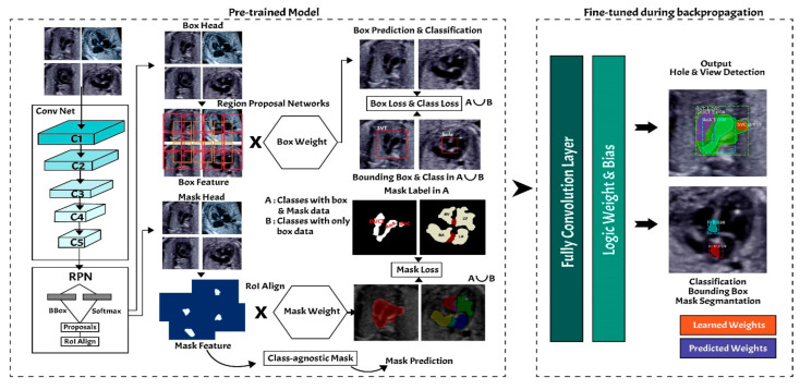
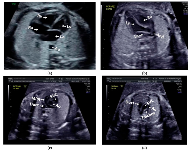
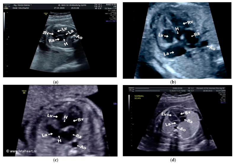
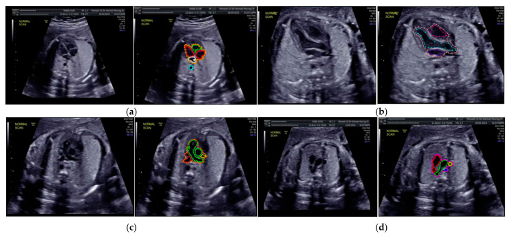
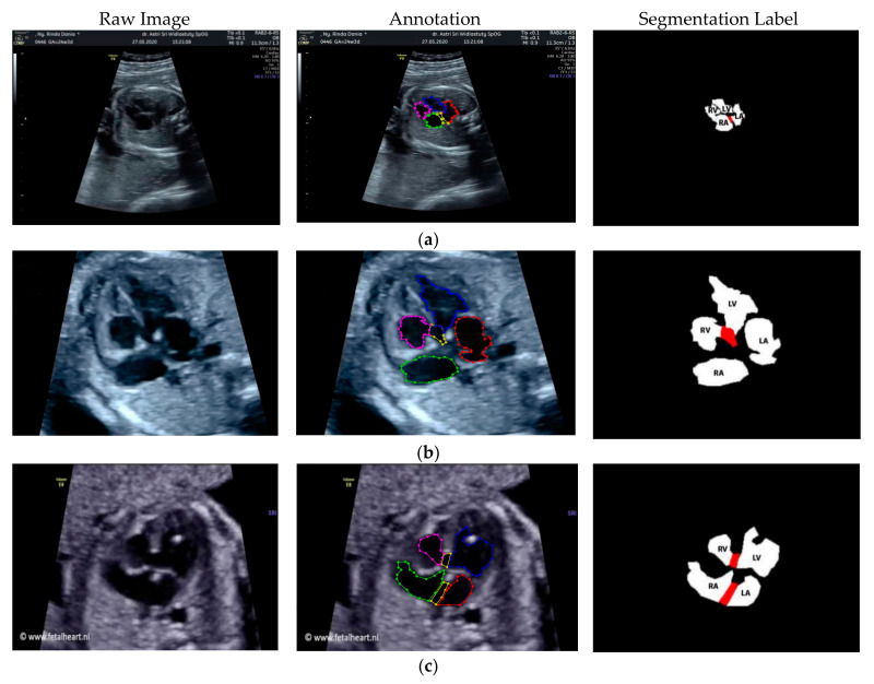
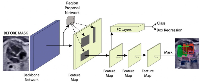
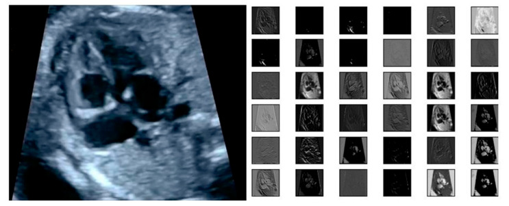
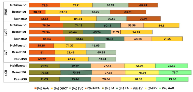
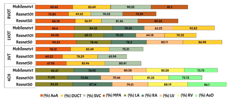
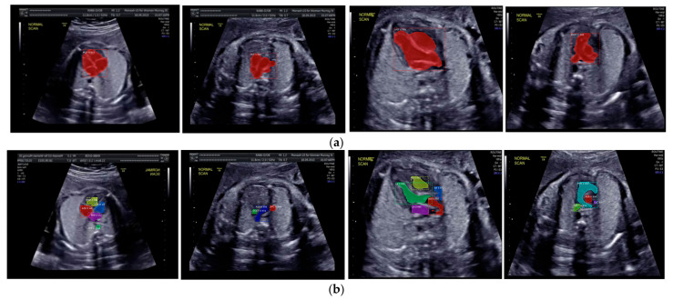
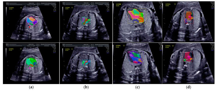
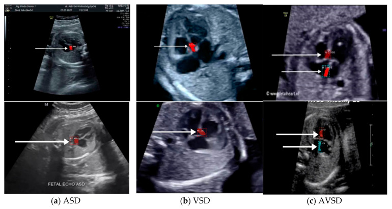
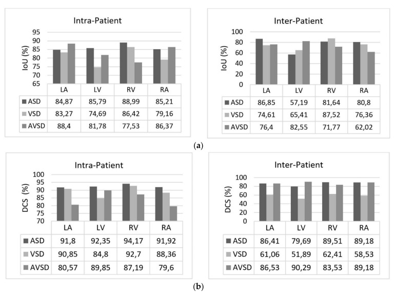
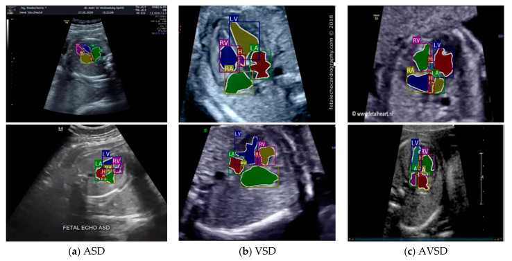
Accurate segmentation of fetal heart in echocardiography images is essential for detecting the structural abnormalities such as congenital heart defects (CHDs). Due to the wide variations attributed to different factors, such as maternal obesity, abdominal scars, amniotic fluid volume, and great vessel connections, this process is still a challenging problem. CHDs detection with expertise in general are substandard; the accuracy of measurements remains highly dependent on humans' training, skills, and experience. To make such a process automatic, this study proposes deep learning-based computer-aided fetal heart echocardiography examinations with an instance segmentation approach, which inherently segments the four standard heart views and detects the defect simultaneously. We conducted several experiments with 1149 fetal heart images for predicting 24 objects, including four shapes of fetal heart standard views, 17 objects of heart-chambers in each view, and three cases of congenital heart defect. The result showed that the proposed model performed satisfactory performance for standard views segmentation, with a 79.97% intersection over union and 89.70% Dice coefficient similarity. It also performed well in the CHDs detection, with mean average precision around 98.30% for intra-patient variation and 82.42% for inter-patient variation. We believe that automatic segmentation and detection techniques could make an important contribution toward improving congenital heart disease diagnosis rates.

Keywords: deep learning; fetal echocardiography; fetal heart standard view; heart defect; instance segmentation.


Conflict of interest statement

The authors declare no conflict of interest.

Figures

Figure 1
Figure 1
The proposes workflow of fetal heart standard view segmentation for heart defect detection with instance segmentation approach.
Figure 2
Figure 2
Fetal heart scan in four standard views of normal anatomy: (a) 4CH; (b) LVOT; (c) RVOT; and (d) 3VT.
Figure 3
Figure 3
Fetal heart scan in 4CH view for CHDs detection: (a) ASD; (b) VSD; (c) AVSD; and (d) Normal.
Figure 4
Figure 4
The sample of annotated images by maternal–fetal clinician for standard fetal heart view segmentation in (a) 4CH (orange: view, cyan: AoA, red: LA, grey: RA, green: LV, and red: RV); (b) LVOT (orange: view, cyan: LA, purple: RV, and blue: LV); (c) RVOT (orange: view, green: MPA, red: DUCT, and yellow: SVC); and (d) 3VT (purple: view, yellow: AoA, green: SVC, and red: DUCT); based on normal anatomy.
Figure 5
Figure 5
The sample of annotated image by maternal–fetal clinician for heart defect detection in case: (a) ASD; (b) VSD; and (c) AVSD. In the annotation, the green line is RA, the red line is LA, the purple line is RV, the blue line is LV, and the yellow line is defect.
Figure 6
Figure 6
Instance segmentation approach.
Figure 7
Figure 7
The example of feature map extracted from ResNet50 architecture in the RPNs back bone.
Figure 8
Figure 8
The IoU performance in heart chamber segmentation in four fetal heart standard views.
Figure 9
Figure 9
The DCS performance in heart chamber segmentation in four fetal heart standard views.
Figure 10
Figure 10
The sample segmentation result of standard view and heart chamber for normal heart anatomy structure: (a) red color contour denotes the fetal heart boundary segmentation in each view, from left to right are 4CH, 3VT, LVOT, and RVOT; (b) heart chamber segmentation in each view from left to right are 4CH (red: RA, purple: LA, yellow: RV, and blue: LV), 3VT (green: DUCT, blue: AoA, and red: SVC), LVOT (green: LV, red: AoA, blue: RA, and yellow: RV), and RVOT (green: DUCT, cyan: MPA, red: AoA, and purple: SVC).
Figure 11
Figure 11
Fetal heart view with heart chamber segmentation in (a) 4CH, (b) 3VT, (c) LVOT, and (d) RVOT for normal heart anatomy structure. Fetal heart view boundary and heart chamber part as the same description with Figure 10.
Figure 12
Figure 12
Sample image result of CHDs detection with 4CH view. The white arrow indicates the defect, whereas red and blue colors are the defect position in the heart septum.
Figure 13
Figure 13
The performance in fetal heart chamber segmentation in 4CH view based on intra- and inter-patient scenario: (a) IoU and (b) DCS.
Figure 14
Figure 14
The sample result of wall-chamber segmentation with 4 CH view in ASD, VSD, and AVSD condition based on abnormal anatomy structure.
Figure 15
Figure 15
Loss curve of heart defect detection with proposed instance segmentation model. RPNs and FCN loss in training and validation set.

References

    1. Bensemlali M., Bajolle F., Laux D., Parisot P., Ladouceur M., Fermont L., Lévy M., Le Bidois J., Raimondi F., Ville Y., et al. Neonatal management and outcomes of prenatally diagnosed CHDs. Cardiol. Young. 2017;27:344–353. doi: 10.1017/S1047951116000639. - DOI - PubMed
    1. Yoon S.A., Hong W.H., Cho H.J. Congenital heart disease diagnosed with echocardiogram in newborns with asymptomatic cardiac murmurs: A systematic review. BMC Pediatr. 2020;20:1–10. doi: 10.1186/s12887-020-02212-8. - DOI - PMC - PubMed
    1. Mcleod G., Shum K., Gupta T., Chakravorty S., Kachur S., Bienvenu L., Shah S.B. Echocardiography in Congenital Heart Disease. Prog. Cardiovasc. Dis. 2018;61:468–475. doi: 10.1016/j.pcad.2018.11.004. - DOI - PubMed
    1. Patel N., Narasimhan E., Kennedy A. Fetal Cardiac US: Techniques and Normal Anatomy Correlated with Adult CT and MR Imaging. RadioGraphics. 2017;37:1290–1303. doi: 10.1148/rg.2017160126. - DOI - PubMed
    1. Puri K., Allen H.D., Qureshi A.M. Congenital Heart Disease. Pediatr. Rev. 2017;38:471–486. doi: 10.1542/pir.2017-0032. - DOI - PubMed