Review
. 2021 Nov 27;22(23):12828.
doi: 10.3390/ijms222312828.

The Role of EREG/EGFR Pathway in Tumor Progression

Affiliations
Review

The Role of EREG/EGFR Pathway in Tumor Progression

Wan-Li Cheng et al. Int J Mol Sci. .

Abstract

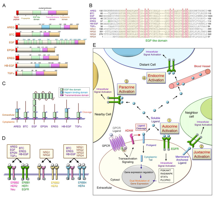
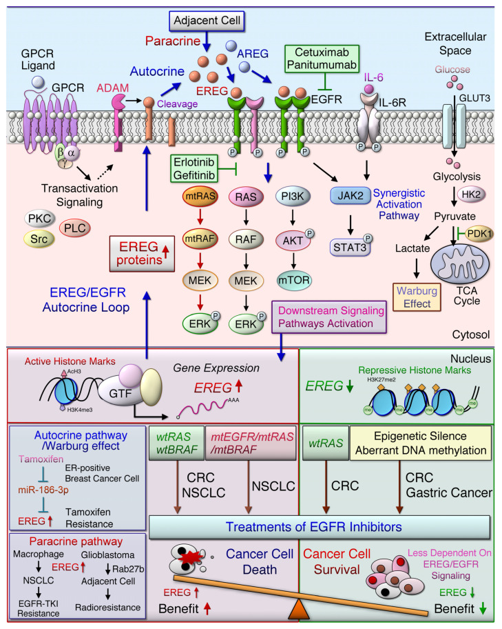
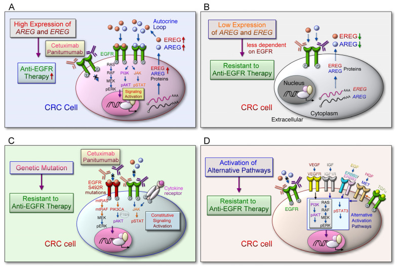
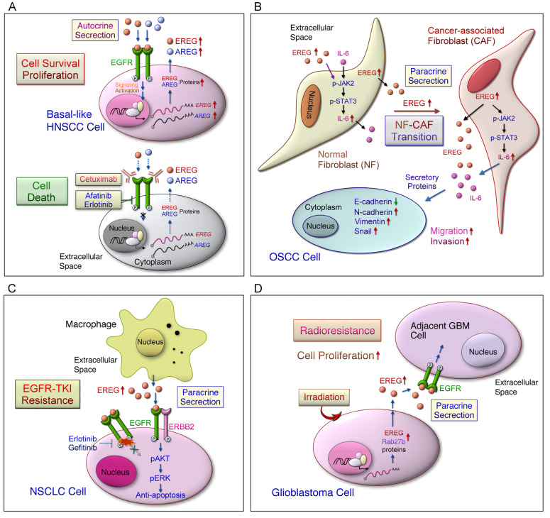
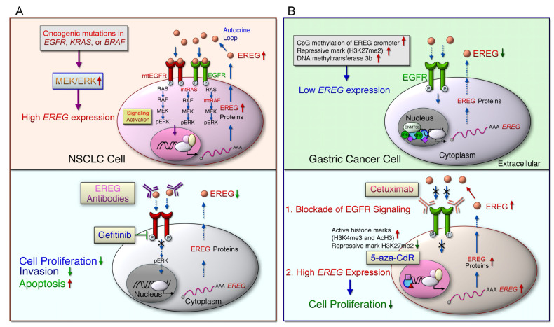
Aberrant activation of the epidermal growth factor receptor (EGFR/ERBB1) by erythroblastic leukemia viral oncogene homolog (ERBB) ligands contributes to various tumor malignancies, including lung cancer and colorectal cancer (CRC). Epiregulin (EREG) is one of the EGFR ligands and is low expressed in most normal tissues. Elevated EREG in various cancers mainly activates EGFR signaling pathways and promotes cancer progression. Notably, a higher EREG expression level in CRC with wild-type Kirsten rat sarcoma viral oncogene homolog (KRAS) is related to better efficacy of therapeutic treatment. By contrast, the resistance of anti-EGFR therapy in CRC was driven by low EREG expression, aberrant genetic mutation and signal pathway alterations. Additionally, EREG overexpression in non-small cell lung cancer (NSCLC) is anticipated to be a therapeutic target for EGFR-tyrosine kinase inhibitor (EGFR-TKI). However, recent findings indicate that EREG derived from macrophages promotes NSCLC cell resistance to EGFR-TKI treatment. The emerging events of EREG-mediated tumor promotion signals are generated by autocrine and paracrine loops that arise from tumor epithelial cells, fibroblasts, and macrophages in the tumor microenvironment (TME). The TME is a crucial element for the development of various cancer types and drug resistance. The regulation of EREG/EGFR pathways depends on distinct oncogenic driver mutations and cell contexts that allows specific pharmacological targeting alone or combinational treatment for tailored therapy. Novel strategies targeting EREG/EGFR, tumor-associated macrophages, and alternative activation oncoproteins are under development or undergoing clinical trials. In this review, we summarize the clinical outcomes of EREG expression and the interaction of this ligand in the TME. The EREG/EGFR pathway may be a potential target and may be combined with other driver mutation targets to combat specific cancers.

Keywords: cancer therapy; epidermal growth factor receptor (EGFR); epiregulin (EREG); tumor microenvironment.


Conflict of interest statement

The authors declare no conflict of interest.

Figures

Figure 1
Figure 1
Protein domains, corresponding receptors of ERBB ligands and the possible activation pathways. (A) Erythroblastic leukemia viral oncogene homolog (ERBB) ligands include a signal peptide, a propeptide region, an epidermal growth factor (EGF)-like domain, a juxtamembrane, a transmembrane, and a cytoplasmic tail. Schematic representation of the membrane-anchored precursor form of the seven human EGF receptor (EGFR) ligands: EGF, transforming growth factor-a (TGFA), heparin-binding EGF-like growth factor (HB-EGF), amphiregulin (AREG), betacellulin (BTC), epiregulin (EREG), and epigen (EPGN). Amino acid residues that constitute the domains in the individual EGFR ligands are listed. EGF consists of nine EGF-like repeats. (B) The yellow region includes aligned amino acid sequences of EGF-like domains in seven EGFR ligands and neuregulin 1-4 (NRG1-4). Asterisks (*) indicate strictly conserved residues. The domains I and III EGFR interacted with the N57 residue of EREG. (C) Arrowheads indicate proximal and distal sites of cleavage in the EGF-like domains, which release to soluble ligands. (D) The ligand binds to the ERBB receptor to form receptor homodimers and heterodimers, and activates the intrinsic kinase domain that recruits proteins to activate intracellular signaling pathways. (E) Soluble ERBB ligands can bind to and activate their receptors (such as EGFR) through endocrine (distant cells), paracrine (adjacent cells), or autocrine (same cell) ways.
Figure 2
Figure 2
The EGFR/ERBB signaling pathway mediated by EREG leads to the cancer development and distinct drug response. G-protein-coupled receptor (GPCR) activation induces the cleavage of transmembrane epiregulin (EREG) protein and then secretes mature EREG. Soluble EREG binds to ERBB, such as epidermal growth factor receptor (EGFR) and ERBB4, which initiate the downstream signaling cascade, whereas the ligand protein is cleaved by a disintegrin and metalloproteinase enzyme (ADAM). The homodimerized or heterodimerized ERBB activate RAS (rat sarcoma)/RAF (rapidly accelerated fibrosarcoma) and phosphatidylinositol-4,5-bisphosphate 3-kinase (PI3K)/AKT (a serine/threonine protein kinase) signaling cascades and synergistically activate, signal transducer and activator of transcription (STAT) 3 signaling pathways, which then induced the upregulation of EREG downstream signaling pathways. Oncogenic mutations in EGFR, KRAS, or BRAF genes in non-small cell lung cancer (NSCLC) cells lead to the constitutive activation of the downstream signaling, which in turn upregulates EREG expression. Treatment with anti-EGFR antibodies, such as cetuximab or panitumumab, in patients with metastatic colorectal cancer (mCRC) with wild-type RAS improved patient outcomes. EREG overexpression was found in wild-type, mutant EGFR (mtEGFR), or mutant BRAF (mtBRAF) NSCLC cells that are sensitive to anti-EREG antibodies or an EGFR-tyrosine kinase inhibitor (EGFR-TKI, gefitinib or erlotinib). EREG might diminish TKI-induced NSCLC cell apoptosis through EGFR/ERBB2 and AKT signaling pathways. However, the low-level expression of AREG and EREG in CRC cells indicates that tumors are less dependent on EGFR, which is particularly prone to cause EGFR inhibitors resistance. Aberrant genetic alterations, including mutant RAS (mtRAS) and mtBRAF in CRC, induce resistance to anti-EGFR therapy. Low EREG expression was caused by aberrant histone modification and DNA methylation in a subset of cancer patients, such as those with gastric cancer, which cause resistance to anti-EGFR therapy. The miR-186-3p/EREG axis as a key regulatory pathway can induce the Warburg effect through EGFR signal activation, thereby increasing the expression of glycolytic genes, including glucose transporter 3 (GLUT3), hexokinase 2 (HK2), and pyruvate dehydrogenase kinase 1 (PDK1) in breast cancer cells resistant to tamoxifen. In addition, Rab27b mediates radioresistance in highly malignant glioblastoma (GBM) cells through the EREG-mediated paracrine pathway.
Figure 3
Figure 3
The alternative pathways and mechanisms bypass targeting EREG-mediated EGFR signal activation in colorectal cancer cells. (A) The expression of AREG and EREG are coordinately regulated by an autocrine loop through EGFR downstream signaling activation, which plays an important role in tumor growth and survival. The EGFR ligand binds to the EGFR and causes downstream signaling pathways that are essential for cell growth and proliferation. Cetuximab or panitumumab prevents the ligand from binding to EGFR, thereby blocking EGFR signaling. (B) Low AREG and EREG gene expression levels are associated with resistance to anti-EGFR therapy. The low expression levels of AREG and EREG indicate that tumor progression is less dependent on EGFR activation; therefore, the cancer cells are particularly prone to less response to EGFR inhibitor treatment. (C) Aberrant genetic alterations, including RAS, BRAF, PIK3CA, EGFR S492R mutations, PTEN loss, and STAT3 phosphorylation in the EGFR signaling pathways induce resistance to anti-EGFR therapy. These constitutively activate the downstream signal cascade of EGFR leading to resistance to anti-EGFR therapy, regardless of EGFR blockade. (D) Aberrant activation of the alternative pathways can induce resistance to anti-EGFR therapy. EGFR downstream effectors can be activated by activating compensatory membrane growth factor receptors, including IGF-1R, MET, HER2 and VEGFR. The stimulation of the corresponding growth factors causes the intracellular signaling pathway to bypass EGFR and induce tumor cell growth and proliferation, leading to resistance to anti-EGFR therapy.
Figure 4
Figure 4
Elevated EREG expression in certain cell types may alter tumorigenesis and therapeutic response in the tumor microenvironment. (A) Inter-tumor heterogeneity may hinder the therapeutic efficiency of anti-EGFR treatments in head and neck squamous cell carcinomas (HNSCC). This may be caused by the dysregulated expression of factors, such as EREG, involved in the EGFR signaling pathway. Notably, basal-like cell lines are more sensitive to EGFR blockade alone or in combination with treatments targeting MEK, mTOR, or ERBB2. Additionally, EREG expression may be a predictive functional marker of anti-EGFR therapy in basal-like HNSCC. (B) The local resident normal fibroblasts (NFs) are converted to cancer-associated fibroblasts (CAFs) in oral squamous cell carcinoma (OSCC), which exhibit tumor-supportive properties. EREG is the most remarkably upregulated gene in CAFs. Overexpression of EREG in NFs activated the CAF phenotype. Mechanistically, the JAK2/STAT3 pathway was enhanced by EREG in parallel with increased IL-6 expression. IL-6 induced the JAK2/STAT3/EREG pathway in a feedback loop. Moreover, EREG-induced CAF activation promotes the epithelial-mesenchymal transition (EMT) necessary for migration and invasion, which depends on JAK2/STAT3 signaling and IL-6. (C) Among EGFR ligands, EREG significantly reduces the sensitivity of cells to EGFR TKI, which may be correlated with the resistance to erlotinib in NSCLC patients. EREG induces AKT phosphorylation in an ERBB2-dependent manner and attenuates TKI-induced apoptosis. Regardless of treatment, EREG induces the formation of EGFR/ERBB2 heterodimers. However, overexpression or knockdown of EREG in cancer cells has little effect on TKI sensitivity. EREG-rich macrophage conditioned medium induces EGFR-TKI resistance. (D) Rab27b mediates radioresistance in highly malignant glioblastoma (GBM) cells. In addition, Rab27b promotes the proliferation of neighboring cells through EREG-mediated paracrine signals after irradiation.
Figure 5
Figure 5
Potential targeting of EREG/EGFR may be applied to a subset of NSCLC and gastric cancer patients. (A) High EREG expression levels were found in the EGFR-mutant, BRAF-mutant NSCLC cells, a subset of NSCLC cells with wild-type EGFR/KRAS/BRAF. In NSCLC cells overexpressing EREG, the inhibition of MEK or ERK could reduce the expression of EREG regardless of the mutation status. Therefore, the activation of the MEK/ERK pathway is a common mechanism of EREG upregulation in NSCLC. EREG levels are decreased by siRNA-mediated EGFR knockdown and EGFR inhibitors in EGFR-mutant NSCLC cells. Moreover, lung tumors of mutant EGFR transgenic mice exhibit high EREG expression. In NSCLC cells with EGFR mutations, both EREG knockdown and anti-EREG antibodies inhibit cell proliferation and invasion and induce apoptosis. Collectively, targeting EREG may be a therapeutic option for EGFR-mutant NSCLC cells with resistance to EGFR-TKIs. (B) EREG is epigenetically silenced in gastric cancer cells through aberrant DNA methylation and histone modification. EREG is methylated and reduced in human gastric cancer cells and primary tissues from a subset of gastric cancer patients. EREG gene expression was reduced by aberrant CpG methylation of the EREG promoter. In addition, treatment with 5-aza-CdR demethylated the CpG site in the EREG promoter, which resulted in the rescue of EREG expression. DNA methyltransferase 3 beta (DNMT3b) predominantly regulates CpG methylation and silencing of the EREG gene. Moreover, treatment with 5-aza-CdR dynamically increased active histone marks (H3K4me3 and AcH3) and decreased the repressive mark (H3K27me2). The combination treatment with 5-aza-CdR and cetuximab exerts a synergistic antiproliferative effect on gastric cancer cells.

References

    1. Jin W. ErBb Family Proteins in Cholangiocarcinoma and Clinical Implications. J. Clin. Med. 2020;9:2255. doi: 10.3390/jcm9072255. - DOI - PMC - PubMed
    1. Yarden Y., Sliwkowski M.X. Untangling the ErbB signalling network. Nat. Rev. Mol. Cell Biol. 2001;2:127–137. doi: 10.1038/35052073. - DOI - PubMed
    1. Yarden Y., Pines G. The ERBB network: At last, cancer therapy meets systems biology. Nat. Rev. Cancer. 2012;12:553–563. doi: 10.1038/nrc3309. - DOI - PubMed
    1. Ledonne A., Mercuri N.B. On the Modulatory Roles of Neuregulins/ErbB Signaling on Synaptic Plasticity. Int. J. Mol. Sci. 2019;21:275. doi: 10.3390/ijms21010275. - DOI - PMC - PubMed
    1. Berdiel-Acer M., Maia A., Hristova Z., Borgoni S., Vetter M., Burmester S., Becki C., Michels B., Abnaof K., Binenbaum I., et al. Stromal NRG1 in luminal breast cancer defines pro-fibrotic and migratory cancer-associated fibroblasts. Oncogene. 2021;40:2651–2666. doi: 10.1038/s41388-021-01719-3. - DOI - PMC - PubMed