. 2021 Nov 30;22(23):13001.
doi: 10.3390/ijms222313001.

Ultrastructural Study of Platelet Behavior and Interrelationship in Sprouting and Intussusceptive Angiogenesis during Arterial Intimal Thickening Formation

Affiliations

Ultrastructural Study of Platelet Behavior and Interrelationship in Sprouting and Intussusceptive Angiogenesis during Arterial Intimal Thickening Formation

Lucio Díaz-Flores et al. Int J Mol Sci. .

Abstract

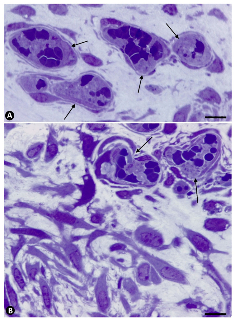
Platelets in atherosclerosis, bypass stenosis, and restenosis have been extensively assessed. However, a sequential ultrastructural study of platelets in angiogenesis during the early phases of these lesions has received less attention. Our objective was the study of platelets in angiogenesis and vessel regression during intimal thickening (IT) formation, a precursor process of these occlusive vascular diseases. For this purpose, we used an experimental model of rat occluded arteries and procedures for ultrastructural observation. The results show (a) the absence of platelet adhesion in the de-endothelialized occluded arterial segment isolated from the circulation, (b) that intraarterial myriad platelets contributed from neovessels originated by sprouting angiogenesis from the periarterial microvasculature, (c) the association of platelets with blood components (fibrin, neutrophils, macrophages, and eosinophils) and non-polarized endothelial cells (ECs) forming aggregates (spheroids) in the arterial lumen, (d) the establishment of peg-and-socket junctions between platelets and polarized Ecs during intussusceptive angiogenesis originated from the EC aggregates, with the initial formation of IT, and (e) the aggregation of platelets in regressing neovessels ('transitory paracrine organoid') and IT increases. In conclusion, in sprouting and intussusceptive angiogenesis and vessel regression during IT formation, we contribute sequential ultrastructural findings on platelet behavior and relationships, which can be the basis for further studies using other procedures.

Keywords: intimal thickening; intussusceptive angiogenesis; peg-and-socket junctions; platelets; sprouting angiogenesis; ultrastructure; vascular regression.


Conflict of interest statement

The authors declare no conflict of interest.

Figures

Figure 1
Figure 1
(AD). Control: Image of an unmodified artery is observed in semithin and ultrathin sections (A,B). Normal microvasculature in the connective and adipose periarterial tissues is also seen (C,D, arrows). A normal vessel and a few interstitial mononuclear cells are present in the periarterial tissues at the end of a sham-operated arterial segment (D). (EH): Initial stage in occluded arteries. De-endothelialized arteries are observed without platelet adhesion. Note the extracellular matrix (asterisk) and absence of adhered platelets or re-endothelialization (E,F). Occasional cell debris are present (F, arrowhead). Absence of platelet adhesion is observed even when neovessels are present in the artery wall (arrows) without crossing the internal elastic membrane of the artery (G,H). Arterial lumen: lu. Adipocytes: ad. Endothelial cell: ec. Smooth muscle cell: smc. (A,C,D) and (G,H): Semithin section. Toluidine blue staining. (B,F): Ultrathin section: Uranyl acetate and lead citrate. Bar: (A,G,H): 15µm. (B): 6µm. (C,D): 20 µm. (E): 10 µm. (F): 2 µm.
Figure 2
Figure 2
Platelets in newly formed vessels that cross the wall of occluded arteries. (A): Semithin section in which numerous platelets, independent of each other, are observed in neovessels (arrows) penetrating the media layer and retained by the internal elastic membrane (iel) of an artery. (B): Ultrastructural image in which a vessel with intraluminal platelets (arrows) crosses the internal elastic lamina (iel), acquiring an hourglass appearance. Endothelial cell: ec. (A): Semithin section. Toluidine blue staining. (B): Ultrathin section: Uranyl acetate and lead citrate: Bar: (A): 15 µm (B): 3.5 µm.
Figure 3
Figure 3
Association of platelets with blood components contributed to the lumen of occluded arteries from the periarterial microvasculature. (A): Relationship of the platelets with fibrinous material and one eosinophil (e). Note the characteristic granules in the latter. (B): Relationship of platelets with a macrophage (m). Arrows point to contacts between platelets and the eosinophil and the macrophage. Ultrathin sections. Uranyl acetate and lead citrate. Bar: (A): 1 µm, (B): 3.5 µm.
Figure 4
Figure 4
Association of platelets with ECs contributed to the lumen of occluded arteries from the periarterial microvasculature. (A): Semithin section in which platelets are observed in association with a portion of an EC aggregate. Note that platelets are present around the aggregate and between the ECs (ec). Insert in (A): A group of platelets at higher magnification. (B): Platelets are seen in the abluminal and luminal surfaces of the ECs that begin their polarization in EC aggregates. This image corresponds to an EC in mitosis (ec). In an abluminal space around ECs presence of membranous material (asterisks). Insert of (B), a detail of the relationship of platelets and another mitotic EC. (A): Semithin section. Toluidine blue staining. (B): Ultrathin section. Uranyl acetate and lead citrate. Bar: (A): 15 µm, (B): 3.5 µm.
Figure 5
Figure 5
Platelets in the abluminal space around ECs. (A): Platelets with a different morphology are observed in association with newly formed basal membrane-like material arranged in multiple layers resembling fingerprints (asterisk). Note a platelet with a projection penetrating an EC (arrow) (compare with peg-and-socket junctions in Figure 6). (B): Presence of granules, similar to those in a modified platelet (arrow), is observed between the membrane-like material in the abluminal space around the EC. Ultrathin sections. Uranyl acetate and lead citrate. Bar: (A,B): 1 µm.
Figure 6
Figure 6
Presence of peg-and-socket junctions between platelets and ECs in the luminal and abluminal EC surfaces. (A): A platelet pseudopod forming a peg is observed penetrating an invagination of the luminal surface of an EC (the socket) (arrow). Insert: A detail at higher magnification of the peg and socket (arrow). (B): A similar peg and socket between a platelet and an EC, but in the abluminal surface of the EC (arrow). Note the fingerprint-like membranous material (asterisk)l around the platelet. The membranous material with or without blood components (e.g., platelets) forms the core, and the surrounding ECs the cover, of the intussusceptive pillars in the arterial lumen (A,B). Ultrathin sections. Uranyl acetate and lead citrate. Bar: (A): 0.7 µm, (B): 0.5 µm.
Figure 7
Figure 7
Platelets during regression of the newly formed vessels. (A): Aggregates of platelets and red blood cells in regressive vessels (arrows). (B): Numerous myointimal cells proliferating around regressive vessels with aggregates of platelets and red blood cells (arrows). Semithin sections. Toluidine blue staining. Bar: (A,B): 15 µm.
Figure 8
Figure 8
Ultrastructural images of aggregates of platelets (arrows) in regressive vessels. Degenerative phenomena are observed in surrounding cells. Ultrathin sections. Uranyl acetate and lead citrate. Bar: (A,B): 0.7 µm.
Figure 9
Figure 9
Numerous extravasated platelets around a regressive vessel plugged by an intraluminal accumulation of red blood cells (asterisk). Note proliferated myointimal cells in the interstitium. (B): Ultrastructural image of extravasated platelets (arrows) around a regressive vessel. Endothelial cell: ec. (A): Semithin section. Toluidine blue staining. (B): Ultrathin section. Uranyl acetate and lead citrate. Bar: (A): 15 µm, (B): 1 µm.
Figure 10
Figure 10
Schematic representation of platelet behavior and interrelationship in occluded arteries. (1) Contribution of platelets and other blood components (with which platelets establish interrelations) to the lumen of the occluded arterial segment, from newly formed vessels, which cross the arterial wall from the periarterial microvasculature, in which they originate by sprouting angiogenesis. (2,3,3A) Incorporation of platelets (colored in blue) between non-polarized ECs that form aggregates (EC spheroids) in the lumen of the artery. (4, 4A) Reorganization of EC aggregates with (a) persistence of platelets in the luminal and abluminal spaces when the ECs are polarized, establishing peg-and-socket junctions with ECs in both spaces (4A, arrow) and (b) platelet participation in the formation of intussusceptive pillar cores together with basal-membrane-like material in the abluminal space (asterisk). (5, 5A) Aggregation of platelets in the lumen of regressive neovessels (‘paracrine transitional organoid’) (arrows) coinciding with myointimal cell proliferation and intimal thickening increase. Formation of a preferential vessel (asterisks). AML: Arterial media layer.

References

    1. Badimon J.J., Ortiz A.F., Meyer B., Mailhac A., Fallon J.T., Falk E., Badimon L., Chesebro J.H., Fuster V. Different response to balloon angioplasty of carotid and coronary arteries: Effects on acute platelet deposition and intimal thickening. Atherosclerosis. 1998;140:307–314. doi: 10.1016/S0021-9150(98)00134-8. - DOI - PubMed
    1. Chandrasekar B., Tanguay J.F. Platelets and restenosis. J. Am. Coll. Cardiol. 2000;35:555–562. doi: 10.1016/S0735-1097(99)00596-3. - DOI - PubMed
    1. Weyrich A.S., Prescott S.M., Zimmerman G.A. Platelets, endothelial cells, inflammatory chemokines, and restenosis: Complex signaling in the vascular play book. Circulation. 2002;106:1433–1435. doi: 10.1161/01.CIR.0000033634.60453.22. - DOI - PubMed
    1. Schulz C., Massberg S. Platelets in atherosclerosis and thrombosis. Handb. Exp. Pharmacol. 2012;210:111–133. - PubMed
    1. Lehle K., Li J., Zimmermann H., Hartmann B., Wehner D., Schmid T., Schmid C. In vitro endothelialization and platelet adhesion on titaniferous upgraded polyether and polycarbonate polyurethanes. Materials. 2014;7:623–636. doi: 10.3390/ma7020623. - DOI - PMC - PubMed

LinkOut - more resources