. 2021 Dec 2;22(23):13034.
doi: 10.3390/ijms222313034.

Human Adenovirus Type 5 Infection Leads to Nuclear Envelope Destabilization and Membrane Permeability Independently of Adenovirus Death Protein

Affiliations

Human Adenovirus Type 5 Infection Leads to Nuclear Envelope Destabilization and Membrane Permeability Independently of Adenovirus Death Protein

Søren Pfitzner et al. Int J Mol Sci. .

Abstract

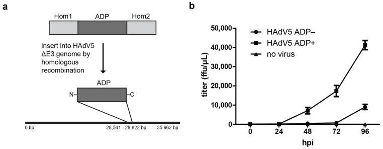
The human adenovirus type 5 (HAdV5) infects epithelial cells of the upper and lower respiratory tract. The virus causes lysis of infected cells and thus enables spread of progeny virions to neighboring cells for the next round of infection. The mechanism of adenovirus virion egress across the nuclear barrier is not known. The human adenovirus death protein (ADP) facilitates the release of virions from infected cells and has been hypothesized to cause membrane damage. Here, we set out to answer whether ADP does indeed increase nuclear membrane damage. We analyzed the nuclear envelope morphology using a combination of fluorescence and state-of-the-art electron microscopy techniques, including serial block-face scanning electron microscopy and electron cryo-tomography of focused ion beam-milled cells. We report multiple destabilization phenotypes of the nuclear envelope in HAdV5 infection. These include reduction of lamin A/C at the nuclear envelope, large-scale membrane invaginations, alterations in double membrane separation distance and small-scale membrane protrusions. Additionally, we measured increased nuclear membrane permeability and detected nuclear envelope lesions under cryoconditions. Unexpectedly, and in contrast to previous hypotheses, ADP did not have an effect on lamin A/C reduction or nuclear permeability.

Keywords: HAdV5; adenovirus death protein; electron cryotomography; membrane damage; nuclear egress; nuclear envelope; serial block-face SEM.


Conflict of interest statement

The authors declare no conflict of interest. The funders had no role in the design of the study; in the collection, analyses, or interpretation of data; in the writing of the manuscript, or in the decision to publish the results.

Figures

Figure 1
Figure 1
ADP expression accelerates HAdV5 particle release from infected cells (a) Overview of ADP insertion into HAdV5 ∆E3 genome by homologous recombination. (b) Growth curve of virus particle release from infected A549 cells from 0 hpi to 96 hpi. The two virus strains HAdV5 ADP- and HAdV5 ADP+ and a control in which no virus was used were compared. The supernatant of infected cells was collected at each time-point and their titer was measured as fluorescence-forming units (ffu)/μL by FACS analysis. Three replicates were measured, and the standard deviation is displayed (n = 3).
Figure 2
Figure 2
HAdV5 infection reduces lamin A/C signal at the nuclear envelope independently of ADP expression (a) A549 cells were infected with HAdV5 ADP− or HAdV5 ADP+ and imaged by live-cell fluorescence microscopy at 48 hpi. The Hoechst 33,342 fluorescence was used to identify infected cells for analysis of their lamin A/C phenotype. A noninfected cell was analyzed in comparison. A representative cell for each phenotype is shown. The dsDNA signal is represented by Hoechst 33,342 stain (Hoechst). The nuclear lamina is represented by an mTagGFP-nanobody recognizing lamin A/C (Lamin A/C). The signal overlap is represented in color (merge). Nuclear membrane regions of interest are enlarged (zoom) with colored corners indicating the channel color. (b) Quantification of anti-lamin A/C mTagGFP-nanobody signal intensity at the nuclear envelope in infection with and without ADP. The fluorescence intensity of multiple cells is shown as a boxplot including the median, upper quartile, lower quartile, maximum and minimum of the population (n = 20). Statistical significance was calculated using a one-way ANOVA with post hoc Tukey test (ns = not significant; *** = p < 0.001).
Figure 3
Figure 3
The nuclear envelope in HAdV5 infection loses overall large-scale stability, and electron cryotomography analyses reveal multiple small-scale instability phenotypes. (a) A549 cells were infected with HAdV5 pV-mCherry ADP+ [27] and fixed at 42 hpi. The pV-mCherry fluorescence was used to identify infected cells for serial block-face SEM sample preparation. A noninfected cell was analyzed in comparison. The 3D volumes of cells of interest were recorded by serial block-face SEM. The nuclear envelopes of a noninfected and infected cell were manually segmented using the IMOD segmentation tool [28]. A mesh overview of both nuclei is shown in images (i) and (ii). An overlay of a surface rendering onto a SEM z-image is shown in images (iii) and (iv). An enlargement of a surface rendering of the nuclear envelope is shown in images (v) and (vi). (b) Electron cryotomograms of multiple membrane modulations observable in HAdV5 pIX-mCherry ADP+ infection. Per phenotype, two example tomograms are shown. Abnormally close contacts between the inner and outer membrane are shown in images (i) and (iv), including a virion detected in the cytoplasm. Outer nuclear membrane protrusions are shown in images (ii) and (v) and double membrane protrusions are shown in images (iii) and (vi). The cytoplasm (cp) and nucleoplasm (np) are labeled.
Figure 4
Figure 4
HAdV5 infection induces small membrane lesions detectable by serial block-face SEM. (a) Examples of nuclear envelope damage detected in infection with HAdV5 pIX-mCherry ADP− and HAdV5 pIX-mCherry ADP+. A549 cells were infected and prepared for serial block-face SEM at 42 hpi. Arrows indicate sites of damage, and the nucleoplasm (np) and cytoplasm (cp) are labeled. (b) Sequence of successive z-slices recorded by serial block-face SEM showing nuclear envelope disruptions by HAdV5 pIX-mCherry ADP− and HAdV5 pIX-mCherry ADP+ infection. A noninfected cell was analyzed for comparison. An overview image for each condition is shown, with a white square indicating a selected ROI. Sequential z-sections of the ROIs are shown, sites of envelope damage are indicated by arrows and the nucleoplasm (np) and cytoplasm (cp) are labeled. A 3D visualization of the membrane damage site of HAdV5 pIX-mCherry ADP+ is shown in Movie S1.
Figure 5
Figure 5
Infection with HAdV5 increases permeability of the nuclear envelope independently of ADP expression. A549 cells were infected with HAdV5 pIX-mCherry ADP− or HAdV5 pIX-mCherry ADP+ and analyzed at 48 hpi. (a) Phenotype of A549 cells after incubation in dextran-FITC. The cells were imaged by live-cell confocal spinning-disk fluorescence microscopy. A representative cell is shown for each condition. The dsDNA signal is represented by Hoechst 33,342 stain (Hoechst). The permeability probe dextran-FITC is detected via the FITC fluorescence (Dextran-FITC). Infected cells were detected by positive expression of pIX-mCherry (pIX-mCherry). The signal overlap is represented in color (merge). (b) Representation of ROIs for dextran-FITC signal analysis. The nuclear ROI was chosen based on the dsDNA signal, the whole-cell ROI was chosen based on the dextran-FITC cell outline and the cytoplasmic ROI was selected as the whole-cell ROI excluding the nucleus ROI. (c) Quantification of dextran-FITC signal ratio between cytoplasm and nucleus of infected cells (n = 50). The ratio is shown as a box plot including the median, upper quartile, lower quartile, maximum and minimum of the population. Statistical significance was calculated using a one-way ANOVA with post hoc Tukey test (ns = not significant; *** = p < 0.001).
Figure 6
Figure 6
Electron cryotomography reveals nuclear envelope lesion and enlarged nuclear pores in HAdV5 infection. Cryotomograms showing (a) a nuclear pore complex (NPC) in an infected cell with a diameter of 81 nm, (b) an enlarged nuclear pore with a diameter of 128 nm and (c) a site of nuclear envelope disruption with a diameter of 222 nm at its widest opening together with virions outside of the nucleus. For each of the three tomograms, a single z-slice of the reconstructed 3D volume is shown in images (i), (iii) and (v). Areas of nucleoplasm (np), cytoplasm (cp), a nuclear pore complex (NPC) and a lipid droplet (ld) are labeled. The scale bar indicates 100 nm and the measured diameter of the opening for each phenotype is indicated. A zoom-in of the nuclear opening (region indicated by black dotted outline) is shown in the top right corner. A region of interest for each tomogram (region indicated by white dotted outline) is shown in images (ii), (iv) and (vi), respectively. The nuclear envelope (cyan), selected HAdV5 particles (magenta) and a lipid droplet (yellow) were manually segmented using the IMOD segmentation tool [28].
Figure 6
Figure 6
Electron cryotomography reveals nuclear envelope lesion and enlarged nuclear pores in HAdV5 infection. Cryotomograms showing (a) a nuclear pore complex (NPC) in an infected cell with a diameter of 81 nm, (b) an enlarged nuclear pore with a diameter of 128 nm and (c) a site of nuclear envelope disruption with a diameter of 222 nm at its widest opening together with virions outside of the nucleus. For each of the three tomograms, a single z-slice of the reconstructed 3D volume is shown in images (i), (iii) and (v). Areas of nucleoplasm (np), cytoplasm (cp), a nuclear pore complex (NPC) and a lipid droplet (ld) are labeled. The scale bar indicates 100 nm and the measured diameter of the opening for each phenotype is indicated. A zoom-in of the nuclear opening (region indicated by black dotted outline) is shown in the top right corner. A region of interest for each tomogram (region indicated by white dotted outline) is shown in images (ii), (iv) and (vi), respectively. The nuclear envelope (cyan), selected HAdV5 particles (magenta) and a lipid droplet (yellow) were manually segmented using the IMOD segmentation tool [28].
Figure 7
Figure 7
Model of nuclear envelope modulations occurring during HAdV5 nuclear egress. The nuclear double membrane is represented as two separate black lines. (I) The noninfected, intact nuclear envelope has a regular, evenly curved appearance. Nuclear lamins (green lines) decorate the inner nuclear membrane and give it stability. (II) During infection, the nuclear lamina is degraded and redistributed, likely resulting in a loss of nuclear envelope stability. Consequently, the envelope loses its regular appearance and the distance between the two nuclear membrane layers is altered. (III) The nuclear envelope develops small lesions through which few virus particles can escape and can be found in the cytoplasm. Most of the virus particles remain within the nucleus. (IV) The small lesions develop into large nuclear membrane breaks. Large quantities of virus and nucleoplasm escape into the cytoplasm (see Figure S3).

References

    1. Tollefson A.E., Ryerse J.S., Scaria A., Hermiston T.W., Wold W.S.M. The E3-11.6-kDa adenovirus death protein (ADP) is required for efficient cell death: Characterization of cells infected with adp mutants. Virology. 1996;220:152–162. doi: 10.1006/viro.1996.0295. - DOI - PubMed
    1. Abou El Hassan M.A.I., van der Meulen-Muileman I., Abbas S., Kruyt F.A.E. Conditionally Replicating Adenoviruses Kill Tumor Cells via a Basic Apoptotic Machinery-Independent Mechanism That Resembles Necrosis-Like Programmed Cell Death. J. Virol. 2004;78:12243–12251. doi: 10.1128/JVI.78.22.12243-12251.2004. - DOI - PMC - PubMed
    1. Weigert M., Binks A., Dowson S., Leung E.Y.L., Athineos D., Yu X., Mullin M., Walton J.B., Orange C., Ennis D., et al. RIPK3 promotes adenovirus type 5 activity. Cell Death Dis. 2017;8:3206. doi: 10.1038/s41419-017-0110-8. - DOI - PMC - PubMed
    1. Raghava S., Giorda K.M., Romano F.B., Heuck A.P., Hebert D.N. The SV40 late protein VP4 is a viroporin that forms pores to disrupt membranes for viral release. PLoS Pathog. 2011;7:e1002116. doi: 10.1371/journal.ppat.1002116. - DOI - PMC - PubMed
    1. Klupp B.G., Granzow H., Mettenleiter T.C. Nuclear Envelope Breakdown Can Substitute for Primary Envelopment-Mediated Nuclear Egress of Herpesviruses. J. Virol. 2011;85:8285–8292. doi: 10.1128/JVI.00741-11. - DOI - PMC - PubMed

Substances

LinkOut - more resources