Review
. 2021 Dec 2;22(23):13059.
doi: 10.3390/ijms222313059.

Mitochondrial Biogenesis in Neurons: How and Where

Affiliations
Review

Mitochondrial Biogenesis in Neurons: How and Where

Carlos Cardanho-Ramos et al. Int J Mol Sci. .

Abstract

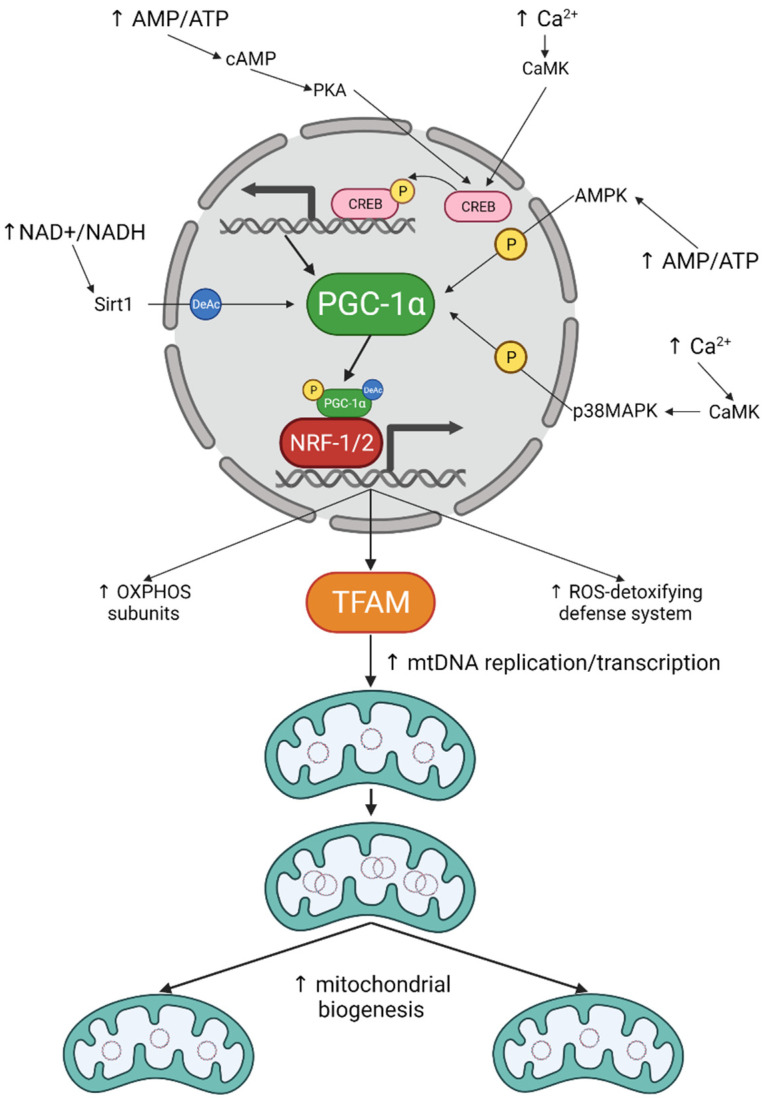
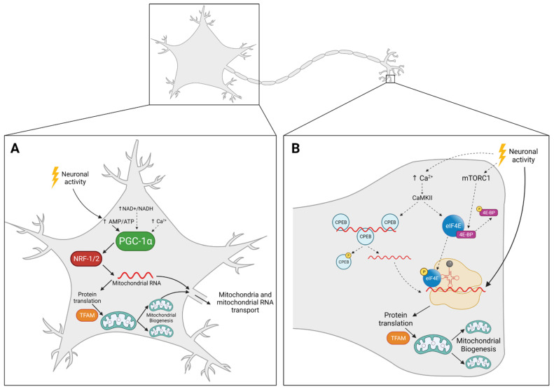
Neurons rely mostly on mitochondria for the production of ATP and Ca2+ homeostasis. As sub-compartmentalized cells, they have different pools of mitochondria in each compartment that are maintained by a constant mitochondrial turnover. It is assumed that most mitochondria are generated in the cell body and then travel to the synapse to exert their functions. Once damaged, mitochondria have to travel back to the cell body for degradation. However, in long cells, like motor neurons, this constant travel back and forth is not an energetically favourable process, thus mitochondrial biogenesis must also occur at the periphery. Ca2+ and ATP levels are the main triggers for mitochondrial biogenesis in the cell body, in a mechanism dependent on the Peroxisome-proliferator-activated γ co-activator-1α-nuclear respiration factors 1 and 2-mitochondrial transcription factor A (PGC-1α-NRF-1/2-TFAM) pathway. However, even though of extreme importance, very little is known about the mechanisms promoting mitochondrial biogenesis away from the cell body. In this review, we bring forward the evoked mechanisms that are at play for mitochondrial biogenesis in the cell body and periphery. Moreover, we postulate that mitochondrial biogenesis may vary locally within the same neuron, and we build upon the hypotheses that, in the periphery, local protein synthesis is responsible for giving all the machinery required for mitochondria to replicate themselves.

Keywords: NRF-1/2; PGC-1α; TFAM; cell body; mitochondrial biogenesis; neurodegenerative diseases; neurons; periphery.


Conflict of interest statement

The authors declare no conflict of interest.

Figures

Figure 1
Figure 1
Mitochondrial biogenesis signalling cascades. Ca2+ and AMP/ATP are the main stimuli for mitochondrial biogenesis. Elevated Ca2+ and AMP levels activate different kinases that can either phosphorylate CREB to promote PGC-1α expression; or directly phosphorylate PGC-1α leading to its activation. Increased NAD+/NADH ratio also results in PGC-1α activation through deacetylation by Sirt1. Once activated, PGC-1α binds to NRF-1/2 to promote expression of several mitochondrial proteins, including TFAM, ultimately resulting in increased mitochondrial biogenesis.
Figure 2
Figure 2
Possible mechanisms for mitochondrial biogenesis in neurons: cell body vs. periphery. (A) Neuronal activity activates PGC-1α through increased AMP/ATP ratio and AMPK. Activation of PGC-1α leads to increased mitochondrial RNA that can either be used for protein translation and mitochondrial renewal or be directly transported to the periphery. Ca2+ and NAD+/NADH ratio may also be involved. (B) Neuronal activity promotes mitochondrial protein translation and mitochondrial biogenesis at the periphery. Although the mechanism is not completely understood, we hypothesized that it may be dependent on Ca2+ and mTORC1. Both Ca2+ and mTORC1 have been seen to increase protein translation in the periphery, whether or not it promotes mitochondrial biogenesis is still unclear. Full arrows represent mechanisms already described in neurons, whereas dashed arrows represent hypothetical mechanisms proposed by the authors.

References

    1. Nunnari J., Suomalainen A. Mitochondria: In Sickness and in Health. Cell. 2012;148:1145–1159. doi: 10.1016/j.cell.2012.02.035. - DOI - PMC - PubMed
    1. Schon E.A., Przedborski S. Mitochondria: The Next (Neurode) Generation. Neuron. 2011;70:1033–1053. doi: 10.1016/j.neuron.2011.06.003. - DOI - PMC - PubMed
    1. Stauch K.L., Purnell P.R., Fox H.S. Quantitative proteomics of synaptic and nonsynaptic mitochondria: Insights for synaptic mitochondrial vulnerability. J. Proteom. Res. 2014;13:2620–2636. doi: 10.1021/pr500295n. - DOI - PMC - PubMed
    1. Völgyi K., Gulyássy P., Háden K., Kis V., Badics K., Kékesi K.A., Simor A., Györffy B., Tóth E.A., Lubec G., et al. Synaptic mitochondria: A brain mitochondria cluster with a specific proteome. J. Proteom. 2015;120:142–157. doi: 10.1016/j.jprot.2015.03.005. - DOI - PubMed
    1. Kiebish M.A., Han X., Cheng H., Luncedord A., Clarke C.F., Moon H., Chuang J.H., Seyfried T.N. Lipidomic analysis and electron transport chain activities in C57BL/6J mouse brain mitchondria. J. Neurochem. 2008;106:299–312. doi: 10.1111/j.1471-4159.2008.05383.x. - DOI - PMC - PubMed

MeSH terms

Substances