. 2021 Dec 3;22(23):13106.
doi: 10.3390/ijms222313106.

Long-Term Effects of Neural Precursor Cell Transplantation on Secondary Injury Processes and Functional Recovery after Severe Cervical Contusion-Compression Spinal Cord Injury

Affiliations

Long-Term Effects of Neural Precursor Cell Transplantation on Secondary Injury Processes and Functional Recovery after Severe Cervical Contusion-Compression Spinal Cord Injury

Alexander Younsi et al. Int J Mol Sci. .

Abstract

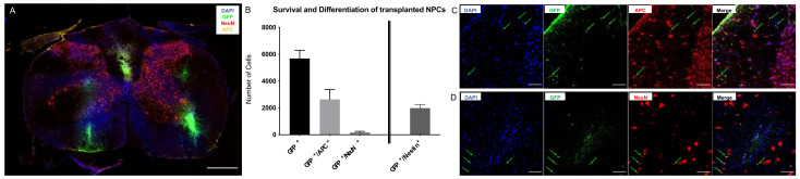
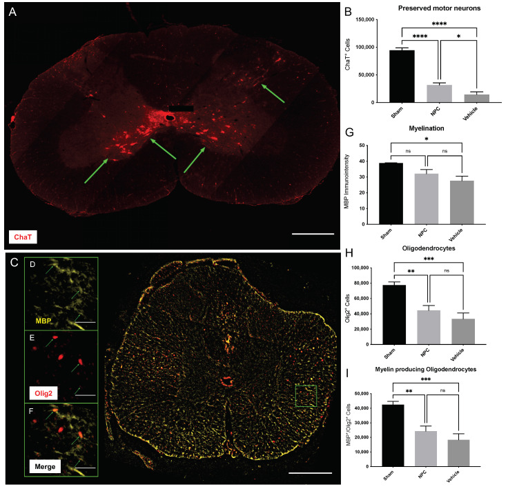
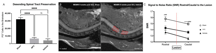
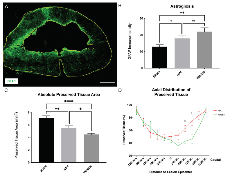
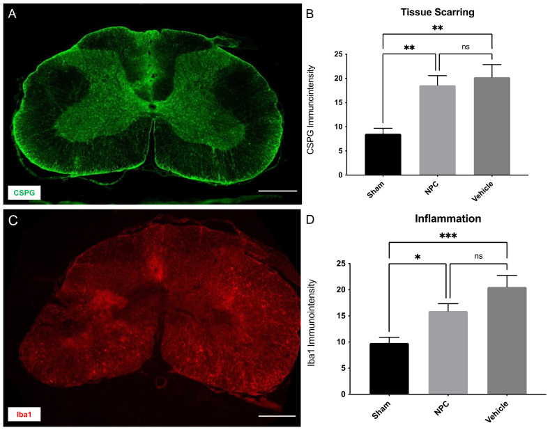
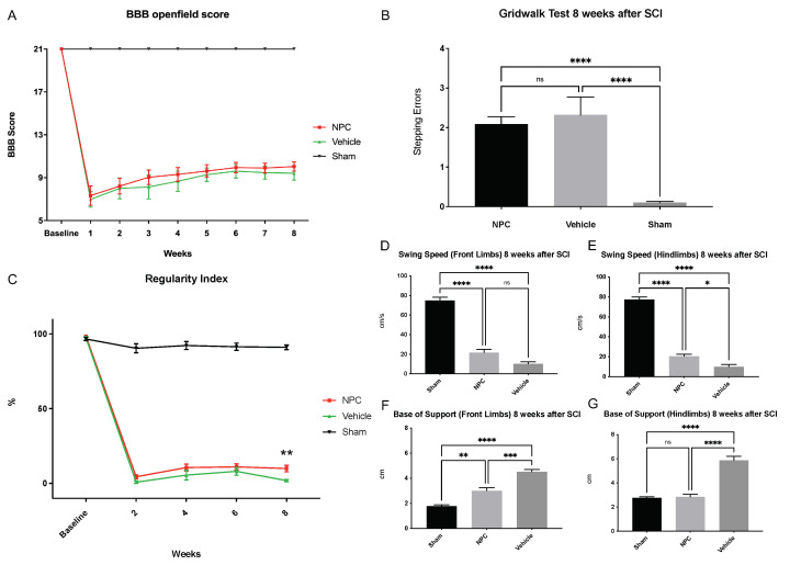
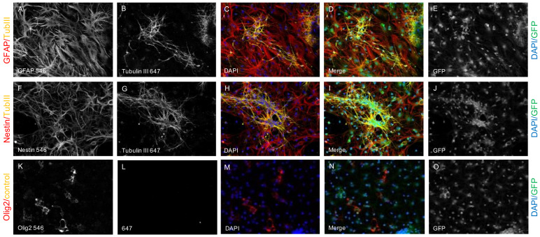
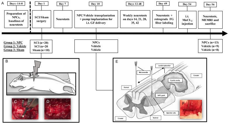
Cervical spinal cord injury (SCI) remains a devastating event without adequate treatment options despite decades of research. In this context, the usefulness of common preclinical SCI models has been criticized. We, therefore, aimed to use a clinically relevant animal model of severe cervical SCI to assess the long-term effects of neural precursor cell (NPC) transplantation on secondary injury processes and functional recovery. To this end, we performed a clip contusion-compression injury at the C6 level in 40 female Wistar rats and a sham surgery in 10 female Wistar rats. NPCs, isolated from the subventricular zone of green fluorescent protein (GFP) expressing transgenic rat embryos, were transplanted ten days after the injury. Functional recovery was assessed weekly, and FluoroGold (FG) retrograde fiber-labeling, as well as manganese-enhanced magnetic resonance imaging (MEMRI), were performed prior to the sacrifice of the animals eight weeks after SCI. After cryosectioning of the spinal cords, immunofluorescence staining was conducted. Results were compared between the treatment groups (NPC, Vehicle, Sham) and statistically analyzed (p < 0.05 was considered significant). Despite the severity of the injury, leading to substantial morbidity and mortality during the experiment, long-term survival of the engrafted NPCs with a predominant differentiation into oligodendrocytes could be observed after eight weeks. While myelination of the injured spinal cord was not significantly improved, NPC treated animals showed a significant increase of intact perilesional motor neurons and preserved spinal tracts compared to untreated Vehicle animals. These findings were associated with enhanced preservation of intact spinal cord tissue. However, reactive astrogliosis and inflammation where not significantly reduced by the NPC-treatment. While differences in the Basso-Beattie-Bresnahan (BBB) score and the Gridwalk test remained insignificant, animals in the NPC group performed significantly better in the more objective CatWalk XT gait analysis, suggesting some beneficial effects of the engrafted NPCs on the functional recovery after severe cervical SCI.

Keywords: NPCs; SCI; functional recovery; neuronal precursor cells; neuroregeneration; spinal cord injury; stem cell therapy.


Conflict of interest statement

The authors declare no conflict of interest. The funders had no role in the design of the study; in the collection, analyses, or interpretation of data; in the writing of the manuscript, or in the decision to publish the results.

Figures

Figure 1
Figure 1
Survival and differentiation of transplanted NPCs eight weeks after severe cervical SCI. (A) Representative image of an NPC animal spinal cord cross-section at 10× magnification depicting the spatial distribution of transplanted NPCs (GFP+/DAPI+), NPC-derived neurons (GFP+/DAPI+/NeuN+) and NPC-derived oligodendrocytes (GFP+/DAPI+/APC+; scale bar = 500 µm). (B) Mean number of surviving NPCs (GFP+), NPC-derived mature neurons (GFP+/NeuN+), NPC-derived mature oligodendrocytes (GFP+/APC+) and undifferentiated NPCs (GFP+/Nestin+) in the injured spinal cord of animals in the NPC group (group 1; n = 8 animals). (C) Colocalization (green arrows) of DAPI, GFP and APC, indicating surviving, viable and mature NPC-derived oligodendrocytes and of (D) DAPI, GFP and NeuN, indicating surviving, viable and mature NPC-derived neurons in an NPC animal at 40× magnification (scale bar = 100 µm).
Figure 2
Figure 2
Spared motor neurons, oligodendrocytes, and myelination in the injured spinal cord eight weeks after severe cervical SCI. (A) Spinal cord cross-section of an NPC animal stained for the motor neuron marker ChaT, indicating spared motor neurons (green arrows) in the ventral grey matter at 10× magnification (scale bar = 500 µm). (B) Significantly more ChaT+ motor neurons could be observed in NPC animals (group 1) compared to Vehicle animals (group 2; n = 8 animals per group; one-way analysis of variance (ANOVA) with post-hoc Tukey-HSD-test; p = 0.0336). (C) Spinal cord cross-section of a Vehicle animal stained for the oligendroglial marker Olig2 and the surrogate marker for myelin MBP at 10× magnification (scale bar = 500 µm). (DF) Enlargement of the green framed inlet in (C), depicting the close spatial relationship (green arrows) of active Olig2+ oligodendrocytes and MBP at 40× magnification (scale bar = 50 µm). (G) The immuno-intensity of the MBP-staining and thus the extent of myelination showed no significant difference between NPC animals and Vehicle animals (n = 8 animals per group; one-way ANOVA with post-hoc Tukey-HSD-test; p = 0.3945). Similarly, the number of Olig2+ oligodendrocytes (H) and Olig2+/MBP+ active oligodendrocytes (I) were not significantly higher in NPC animals compared to Vehicle animals (both n = 8 animals per group; one-way ANOVA with post-hoc Tukey-HSD-test; p = 04549) (* p < 0.05; ** p < 0.01; *** p < 0.001; **** p < 0.0001 and ns = not significant).
Figure 3
Figure 3
Preservation or regeneration of descending and ascending spinal tracts in the injured spinal cord eight weeks after severe SCI. (A) The number of FG+ neurons in the brainstem and thus possibly the extent of descending reticulospinal tract preservation or regeneration was not significantly different between NPC animals (group 1) and Vehicle animals (group 2; n = 5 animals per group; one-way ANOVA with post-hoc Tukey-HSD-test; p = 0.6452). (B) On the left side, sagittal T1-weighted MEMRI image of an uninjured Sham animal (group 3) eight weeks after SCI and 48 h after intrathecal injection of MnCl2, showing faint but nearly homogeneous contrast enhancement of the cervicothoracic spinal cord. On the right side, sagittal T1-weighted MEMRI image of an injured Vehicle animal (group 2) depicting the lesion as a round, hypointense formation in the cervical spinal cord with suspected caudal decrease of Mn2+ signal intensity. Red ROIs depicting the axial slices from 4 mm rostral to 4 mm caudal to the lesion used for detailed SNR measurements. (C) Rostral to caudal decrease of SNR over the lesion was only significant in the Vehicle group (n = 5 animals, n = 4 rostral and n = 4 caudal axial slices per animal; unpaired two-sample t-test, p = 0.007). Correspondingly, SNR caudal to the lesion was significantly lower in Vehicle animals compared to NPC and Sham animals (n = 5 animals per group, n = 4 caudal axial slices per animal; one-way ANOVA with Tukey-HSD-test; p = 0.0017 and p < 0.0001, respectively), indicating more preserved or regenerated descending as well as ascending spinal tracts after NPC-transplantation (* p < 0.05, NPC vs. Sham; *** p < 0.001, NPC vs. Sham; **** p < 0.0001, Vehicle vs. Sham and NPC vs. Sham; ### p < 0.01, Vehicle vs. Sham; #### p < 0.0001, Vehicle vs. Sham; †† p < 0.001, NPC vs. Vehicle; ns = not significant).
Figure 4
Figure 4
Reactive astrogliosis and tissue preservation in the injured spinal cord eight weeks after severe cervical SCI. (A) Spinal cord cross-section of a Vehicle animal stained for GFAP, a marker for reactive astrocytes, at 10× magnification with the entire spinal cord as well as the intramedullary cystic cavity outlined (yellow lines; scale bar = 500 µm). (B) The immuno-intensity of the GFAP-staining and thus the reactive astrogliosis was comparable in NPC animals (group 1) and Vehicle animals (group 2; n = 8 animals per group; one-way ANOVA with Tukey-HSD-test; p = 0.2624). (C) The overall preserved tissue area was significantly higher in NPC animals compared to Vehicle animals (n = 8 animals per group; one-way ANOVA with Tukey-HSD-test; p = 0.043) (D) The percentage of preserved spinal cord tissue along the spinal axis was lowest +/−240 µm from the lesion epicenter in all injured animals, with an increase to 90% towards +/−1200 µm. In comparison to Vehicle animals, NPC animals showed a significant increase of preserved tissue 480 µm and 720 µm caudal from the lesion epicenter (n = 8 animals per group; one-way ANOVA with Tukey-HSD-test; p = 0.0071 and p = 0.0177, respectively) (* p < 0.05; ** p < 0.01; **** p < 0.0001 and ns = not significant).
Figure 5
Figure 5
Tissue scarring and inflammation in the injured spinal cord eight weeks after severe cervical SCI. (A) Spinal cord cross-section of an NPC animal stained for CSPG, a marker for proteoglycans in the extracellular matrix at 10× magnification (scale bar = 500 µm). (B) The immuno-intensity of the CSPG-staining and thus the extent of tissue scarring showed no significant difference between NPC animals (group 1) and Vehicle animals (group 2; n = 8 animals per group; one-way ANOVA with Tukey-HSD-test; p = 0.8281). (C) Spinal cord cross-section of a Vehicle animal stained for Iba1, a marker for macrophages at 10× magnification (scale bar = 500 µm). (D) The immuno-intensity of the Iba1-staining and thus the number of Iba1+ macrophages was not significantly reduced in NPC animals compared to Vehicle animals (n = 8 animals per group; one-way ANOVA with Tukey-HSD-test; p = 0.1468) (* p < 0.05; ** p < 0.01; *** p < 0.001 and ns = not significant).
Figure 6
Figure 6
Functional recovery from baseline until eight weeks after severe cervical SCI. (A) While Sham animals (group 3; n = 8 animals) received 21 points on the BBB score at every timepoint, no significant difference in the BBB score could be observed between NPC animals (group 1) and Vehicle animals (group 2) throughout the experiment and recovery of injured animals generally remained low (n = 13 animals in the NPC group and n = 9 animals in the Vehicle group; two-way repeated measures ANOVA with Tukey test for multiple comparisons). (B) Similarly, assessment of fine motor control with the Gridwalk test revealed no significant difference between the NPC and the Vehicle animals at the end of the experiment (n = 13 NPC animals, n = 9 Vehicle animals and n = 8 Sham animals; one-way ANOVA with Tukey-HSD-test; p = 0.7984). (C) In contrast, the “Regularity Index” of the more objective CatWalk XT automated quantitative gait analysis as a measurement for coordination was significantly improved in NPC animals compared to Vehicle animals eight weeks after the injury (n = 13 NPC animals, n = 9 Vehicle animals and n = 8 Sham animals; two-way repeated measures ANOVA with Tukey test for multiple comparisons; p = 0.0051). Furthermore, while indifferent for the front limbs (D), the “Swing Speed” of the hindlimbs (E) in the CatWalk XT automated quantitative gait analysis was significantly improved in NPC animals compared to Vehicle animals eight weeks after SCI, suggesting less limping after NPC-transplantation (n = 13 NPC animals, n = 9. Vehicle animals and n = 8 Sham animals; Kruskal–Wallis test followed by Dunn’s multiple comparisons test; p = 0.0959 and p = 0.0158, respectively). Similarly, the “Base of Support” indicating trunk stability and weight-bearing when decreased was significantly lower in NPC animals compared to Vehicle animals at the end of the experiment in both, the front limbs (F) and the hindlimbs (G); n = 13 NPC animals, n = 9 Vehicle animals and n = 8 Sham; Kruskal–Wallis test followed by Dunn’s multiple comparisons test; p = 0.0006 and p < 0.0001, respectively) (* p < 0.05; ** p < 0.01; *** p < 0.001; **** p < 0.0001 and ns = not significant).
Figure 7
Figure 7
DAPI+ (blue) and GFP+ (green) cells are expressing Nestin ((F), red) and are thus identified as viable NPCs ((I), green/blue/red). The tripotential differentiation capacity of the NPCs is assessed by costaining of GFP ((E,J,O,D,I,N), green) and DAPI ((C,H,M,D,I,N), blue) with TubIII ((B,G,C,H,D,I), yellow), GFAP ((A,C,D), red) and Olig2 ((K,M,N), red) indicating viable NPC-derived neurons ((D,I), green/blue/yellow), viable NPC-derived astrocytes ((D,I), green/blue/red) and viable NPC-derived oligodendrocytes ((N), green/blue/red). respectively. Only the secondary antibody (Alexa-647) has been used as a negative control (L).
Figure 8
Figure 8
(A) Timeline of the study interventions (e.g., pump implantation for i.t. growth factor (GF) delivery at day 10 or manganese-enhanced magnetic resonance imaging (MEMRI) at day 56), also indicating the group design and the group sizes at the beginning and at the end of the experiment. (B) The contusion-compression SCI is performed at day 1 with a modified aneurysm clip (C), leaving a visible injury on the exposed cervical spinal cord at the C6 level (D). (E) Modified after (47)—NPCs are transplanted into the spinal cord at four distinct sites via stereotactic injection (F) at day 10.

Similar articles

Cited by

References

    1. Furlan J.C., Sakakibara B.M., Miller W.C., Krassioukov A.V. Global incidence and prevalence of traumatic spinal cord injury. Can. J. Neurol. Sci. 2013;40:456–464. doi: 10.1017/S0317167100014530. - DOI - PubMed
    1. Sekhon L.H., Fehlings M.G. Epidemiology, demographics, and pathophysiology of acute spinal cord injury. Spine. 2001;26:S2–S12. doi: 10.1097/00007632-200112151-00002. - DOI - PubMed
    1. Furlan J.C., Fehlings M.G. The impact of age on mortality, impairment, and disability among adults with acute traumatic spinal cord injury. J. Neurotrauma. 2009;26:1707–1717. doi: 10.1089/neu.2009.0888. - DOI - PMC - PubMed
    1. Anderson K.D. Targeting recovery: Priorities of the spinal cord-injured population. J. Neurotrauma. 2004;21:1371–1383. doi: 10.1089/neu.2004.21.1371. - DOI - PubMed
    1. Anderson K.D., Sharp K.G., Steward O. Bilateral cervical contusion spinal cord injury in rats. Exp. Neurol. 2009;220:9–22. doi: 10.1016/j.expneurol.2009.06.012. - DOI - PMC - PubMed

MeSH terms

Substances