. 2021 Nov 25;13(23):5941.
doi: 10.3390/cancers13235941.

Bridging the Species Gap: Morphological and Molecular Comparison of Feline and Human Intestinal Carcinomas

Affiliations

Bridging the Species Gap: Morphological and Molecular Comparison of Feline and Human Intestinal Carcinomas

Tanja Groll et al. Cancers (Basel). .

Abstract

Limited availability of in vivo experimental models for invasive colorectal cancer (CRC) including metastasis and high tumor budding activity is a major problem in colorectal cancer research. In order to compare feline and human intestinal carcinomas, tumors of 49 cats were histologically subtyped, graded and further characterized according to the human WHO classification. Subsequently, feline tumors were compared to a cohort of 1004 human CRC cases. Feline intestinal tumors closely resembled the human phenotype on a histomorphological level. In both species, adenocarcinoma not otherwise specified (ANOS) was the most common WHO subtype. In cats, the second most common subtype of the colon (36.4%), serrated adenocarcinoma (SAC), was overrepresented compared to human CRC (8.7%). Mucinous adenocarcinoma (MAC) was the second most common subtype of the small intestine (12.5%). Intriguingly, feline carcinomas, particularly small intestinal, were generally of high tumor budding (Bd) status (Bd3), which is designated an independent prognostic key factor in human CRC. We also investigated the relevance of feline CTNNB1 exon 2 alterations by Sanger sequencing. In four cases of feline colonic malignancies (3 ANOS, 1 SAC), somatic missense mutations of feline CTNNB1 (p.D32G, p.D32N, p.G34R, and p.S37F) were detected, indicating that mutational alterations of the WNT/β-catenin signaling pathway potentially play an essential role in feline intestinal tumorigenesis comparable to humans and dogs. These results indicate that spontaneous intestinal tumors of cats constitute a useful but so far underutilized model for human CRC. Our study provides a solid foundation for advanced comparative oncology studies and emphasizes the need for further (molecular) characterization of feline intestinal carcinomas.

Keywords: CTNNB1; colorectal cancer; comparative oncology; spontaneous feline intestinal tumors; tumor budding.


Conflict of interest statement

LABOKLIN GmbH & Co. KG offers histopathological service for routine diagnostics. T.G. presented preliminary parts of this study at the 4th Cutting Edge Pathology Congress 2021. W.W. has attended Advisory Boards and served as speaker for Roche, MSD, BMS, AstraZeneca, Pfizer, Merck, Lilly, Boehringer, Novartis, Takeda, Bayer, Amgen, Astellas, Eisai, Illumina, Siemens, Agilent, ADC, GSK, and Molecular Health. W.W. receives research funding from Roche, MSD, BMS and AstraZeneca. N.P. has attended Advisory Boards and served as speaker for Roche, BMS, AstraZeneca, Lilly, Novartis, Bayer, Illumina, and Thermo Fisher Scientific. K.S. is consultant for Roche Pharma AG, member of the advisory board of TRIMT GmbH and has filed a patent on a radiopharmaceutical. All other authors declare no conflicts of interest.

Figures

Figure 1
Figure 1
Tumor budding (TB) in the invasive front of feline intestinal carcinomas according to the 3-tier-system for budding assessment of human CRC (left: Pan-cytokeratin, right: H&E, consecutive sections, 20×). Tumor buds are indicated by arrow heads; (A) Low TB activity (Bd1, Pan-cytokeratin); (B) Low TB activity (Bd1, H&E); (C) Moderate TB activity (Bd2, Pan-cytokeratin); (D) Moderate TB activity (Bd2, H&E); (E) High TB activity (Bd3, Pan-cytokeratin); and (F) High TB activity (Bd3, H&E). For corresponding human H&E sections see Figure S1.
Figure 2
Figure 2
Scoring for nuclear translocated β-catenin in feline intestinal carcinomas (Anti-β-catenin; 20×). (A) Tumor with no or scattered (<5%) nuclear β-catenin staining (score 0); (B) 5–25% of tumor cells display nuclear positivity for β-catenin (score 1); (C) 26–50% of tumor cells display nuclear positivity (score 2); and (D) >50% of tumor cells are positive for nuclear β-catenin (score 3). For corresponding human β-catenin stainings see Figure S2.
Figure 3
Figure 3
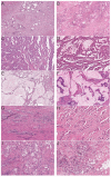
Feline intestinal carcinomas (left) closely resemble human WHO subtypes (right) (H&E). (A) Feline colonic adenocarcinoma not otherwise specified (ANOS, 8×); (B) Human colonic ANOS (8×); (C) Feline colonic serrated adenocarcinoma (SAC, 8×); (D) Human colonic SAC (8×); (E) Feline small intestinal mucinous adenocarcinoma (MAC, 8×); (F) Human colonic MAC (8×); (G) Feline small intestinal micropapillary carcinoma (MPC, 8×); (H) Human colonic MPC (8×); (I) Feline colonic signet-ring cell carcinoma (SRCC, 20×); and (J) Human colonic SRCC (20×).
Figure 4
Figure 4
Characteristic malignancy features of feline intestinal tumors (H&E). (A) Vascular invasion (10×); (B) Lymphangiosis carcinomatosa (4×); (C) Perineural invasion (10×); (D) Lymph node metastasis (4×); (E) Serosal invasion, arrow (1×); and (F) Mucosal ulceration (4×).
Figure 5
Figure 5
Kruskal–Wallis test of WHO subtypes regarding invasiveness. For the overall cohort, statistical analysis revealed a significant difference between the WHO subtypes regarding the feature invasiveness, represented by a cumulative score of invasiveness (vascular (1), perineural (1), lymphatic (1) and serosal invasion (1), max. score of 4). (p = 0.021). The score of invasiveness was significantly higher for ANOS than for SAC (* p = 0.014).
Figure 6
Figure 6
Four distinct somatic missense mutations were detected, each of them in a different case. DNA forward sequences; green: wildtype sequence of normal intestinal tissue (n = 3); red: tumor Table S4. (A) Cytosine (C) is substituted by Thymine (T), resulting in a missense mutation leading to a replacement of Serine (S) by Phenylalanine (F) on the protein level (colonic ANOS; β-catenin score 1); (B) Adenosine (A) is substituted by Guanin (G) resulting in a missense mutation leading to a replacement of Aspartic acid (D) by Glycine (G) (colonic ANOS; score 3). For case 20, no normal tissue was available; (C) Guanin (G) is substituted by Adenosine (A) resulting in a replacement of Aspartic acid (D) by Asparagine (N) (colonic ANOS, score 1); (D) Guanin (G) is substituted by Adenosine (A) resulting in a replacement of Glycine (G) by Arginine (R) (colonic SAC, score 1).

References

    1. Sung H., Ferlay J., Siegel R.L., Laversanne M., Soerjomataram I., Jemal A., Bray F. Global Cancer Statistics 2020: GLOBOCAN Estimates of Incidence and Mortality Worldwide for 36 Cancers in 185 Countries. CA Cancer J. Clin. 2021;71:209–249. doi: 10.3322/caac.21660. - DOI - PubMed
    1. Washington M.K., Powell A.E., Sullivan R., Sundberg J.P., Wright N., Coffey R.J., Dove W.F. Pathology of rodent models of intestinal cancer: Progress report and recommendations. Gastroenterology. 2013;144:705–717. doi: 10.1053/j.gastro.2013.01.067. - DOI - PMC - PubMed
    1. Jackstadt R., Sansom O.J. Mouse models of intestinal cancer. J. Pathol. 2016;238:141–151. doi: 10.1002/path.4645. - DOI - PMC - PubMed
    1. Taketo M.M., Edelmann W. Mouse models of colon cancer. Gastroenterology. 2009;136:780–798. doi: 10.1053/j.gastro.2008.12.049. - DOI - PubMed
    1. McIntyre R.E., Buczacki S.J., Arends M.J., Adams D.J. Mouse models of colorectal cancer as preclinical models. Bioessays. 2015;37:909–920. doi: 10.1002/bies.201500032. - DOI - PMC - PubMed

LinkOut - more resources