Review
. 2021 Nov 26;13(23):5949.
doi: 10.3390/cancers13235949.

Genome Instability in Multiple Myeloma: Facts and Factors

Affiliations
Review

Genome Instability in Multiple Myeloma: Facts and Factors

Anna Y Aksenova et al. Cancers (Basel). .

Abstract

Multiple myeloma (MM) is a malignant neoplasm of terminally differentiated immunoglobulin-producing B lymphocytes called plasma cells. MM is the second most common hematologic malignancy, and it poses a heavy economic and social burden because it remains incurable and confers a profound disability to patients. Despite current progress in MM treatment, the disease invariably recurs, even after the transplantation of autologous hematopoietic stem cells (ASCT). Biological processes leading to a pathological myeloma clone and the mechanisms of further evolution of the disease are far from complete understanding. Genetically, MM is a complex disease that demonstrates a high level of heterogeneity. Myeloma genomes carry numerous genetic changes, including structural genome variations and chromosomal gains and losses, and these changes occur in combinations with point mutations affecting various cellular pathways, including genome maintenance. MM genome instability in its extreme is manifested in mutation kataegis and complex genomic rearrangements: chromothripsis, templated insertions, and chromoplexy. Chemotherapeutic agents used to treat MM add another level of complexity because many of them exacerbate genome instability. Genome abnormalities are driver events and deciphering their mechanisms will help understand the causes of MM and play a pivotal role in developing new therapies.

Keywords: DNA repair; chromothripsis; editing deaminases; genome instability; kataegis; multiple myeloma; translocations.


Conflict of interest statement

The authors declare no conflict of interest.

Figures

Figure 1
Figure 1
Stages of MM development and their characteristics. MM is almost always preceded by a precancerous condition termed monoclonal gammopathy of undetermined significance, MGUS [5,6,7]. Smoldering multiple myeloma (SMM) is an intermediate stage between MGUS and MM [12,13]. MGUS is diagnosed when serum monoclonal protein (M-protein) is detected, but levels are lower than in MM; a 2–3-fold elevation of the number of clonal bone marrow plasma cells is usually found in MGUS compared to healthy individuals. In MGUS, end-organ damage, CRAB, attributed to the plasma cell proliferative disorder, is absent [1]. SMM is diagnosed when serum or urinary monoclonal protein rises and/or the number of clonal bone marrow plasma cells increases, but there is no MDE or amyloidosis [1]. There are germline risk alleles in several genes associated with familiar cases of MM and MGUS; also, numerous SNPs found in GWAS are associated with MM development risk. Telomere status is another factor that can affect MM development. Primary events can be found as early as MGUS and are represented by structural genome changes, gain of chromosomes, and mutations. The primary structural genomic changes are nearly equally represented by the two major groups: the IGH translocations and trisomies of several odd chromosomes (referred to as hyperdiploidy). Few other structural aberrations found as early as the MGUS stage are listed. While the disease progresses, more genomic changes accumulate. The events that are characteristic of SMM and MM, but not MGUS, are classified as secondary. They are represented by various structural genome changes including MYC translocations, accumulation of complex genome rearrangements (partially, since some of them can be seen at MGUS stage), mutations in various pathways, and AID/APOBEC-induced mutation burden. The endpoint of the scheme illustrates treatment-induced events. Some of them can modulate DNA repair and impact genome stability and mutation accumulation. Abbreviations: IMiDs—Immunomodulatory drugs; CRAB—an acronym for Calcium (elevated), Renal failure, Anemia, and Bone lesions, the most common symptoms of MM; MDE—myeloma-defining events (see text for more details); MRI—magnetic resonance imaging; BM—bone marrow; AID—activation-induced cytosine deaminase essential for class switch recombination (CSR) and somatic hypermutation (SHM); APOBEC—a family of cytosine deaminases that play an essential role in mutagenesis in cancer cells; GWAS—Genome-Wide Association Studies.
Figure 2
Figure 2
MM treatment outlines. The schemes align with the latest recommendations and studies and include available options for MM treatment as well as standards of care [87,88,90,91,103]. Action of some drugs on DNA integrity or pathways affecting DNA metabolism/repair is shown on the scheme. All patients can be subdivided into three categories based on frailty: “go-go”, “slow-go”, and “no-go” [111]. “Slow-go” and “no-go” categories are usually treated using the reduced intensity regimens or dose-adjusted regimens and are not shown on the scheme. Treatment for the “no-go” category excludes chemotherapeutic drugs and includes agents with low toxicity, mostly for palliative care [111]. The “go-go” category and to some extent the “slow-go” category are shown on the scheme. VRd * can be used as induction (initial) therapy for newly diagnosed and transplant-eligible patients with MM and is associated with a progression-free survival longer than 4 years and overall survival at 4 years higher than 80% [87,103]. The alternative scheme is VTd, which is slightly less efficient [103]. The addition of the monoclonal antibody daratumumab targeting CD38 to the standard VRd or VTd regimen (Quadruplet regimen, Dara-VRd *, or Dara-VTd) is recommended for high-risk patients [87]. DARA-Rd is also an important alternative to VRd in newly diagnosed MM [112]. Besides that, VCd can be used as an induction scheme [88]. High-dose melphalan and ASCT give a good response and provide the longest remission [87,102]. Single-agent lenalidomide, administered continuously until disease progression is considered the standard of care for maintenance, and bortezomib can be used for high-risk patients. In addition, ixazomib can be considered in place of bortezomib [87,103,113]. If the disease relapses, another ASCT can be tried for eligible patients [100,101,102,114,115]; new treatment regimens are usually considered, including quadruplet schemes and new drugs [87]. The alkylating agent cyclophosphamide can be used instead of immunomodulatory agents as treatment for refractory patients and in patients with renal dysfunction in combination with bortezomib and dexamethasone; this setting can also be used for induction therapy in ASCT-eligible patients [87,116,117,118,119,120,121,122,123,124,125]. Melphalan and prednisone-based therapy$ can be used for ASCT-ineligible patients and often is represented by VMP or DARA-VMP schemes [88]. Continuous therapy or initial therapy with maintenance can be used in a non-transplant setting [113]. A multi-regimen, including bortezomib, an immunomodulatory drug, dexamethasone, cytotoxic cisplatin, doxorubicin, cyclophosphamide, and etoposide, may be used for plasma cell leukemia or extramedullary disease [87]. Schemes that can be used for refractory to lenalidomide and/or bortezomib patients are also given in the center of the figure. Abbreviations: V—bortezomib, DARA—daratumumab, R—lenalidomide, T—thalidomide, POM —pomalidomide, d—dexamethasone; K—carfilzomib, I—ixazomib, E—elotuzumab, Isa—isatuximab, C—cyclophosphamide, P—prednisone; S—selinexor, Ven—venetoclax, ICL—intermolecular crosslinks in the DNA, FA/BRCA—pathway responsible for the repair of intermolecular crosslinks in the DNA, HR—homologous recombination, HRR—homologous recombination repair, DDR—DNA damage response, NER—nucleotide excision repair. *—Lenalidomide-containing regimens (e.g., VRd and Dara-VRd) are not yet approved by European Medicines Agency (EMA, EU) as induction for ASCT-eligible patients, although they offer good risk–benefit profiles and are widely used in the USA [87,88]. **—Bortezomib and ixazomib have not yet been approved by the EMA for maintenance after ASCT [88,113]. ***—Awaiting EMA approval. $—melphalan-containing regimens in this setting are not recommended in the USA due to concerns about SPMs and stem cells damage.
Figure 3
Figure 3
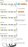
A simplified scheme of B-cell development based on studies in humans and mice. The scheme illustrates the principal stages of B-cell development (predominantly of the B2 lineage) based on studies in mice and humans. Commitment to the B-cell lineage requires the action of several transcription factors (some of them are illustrated in the figure). PAX5, BACH2, PU.1, and IRF8 are expressed throughout B-cell development and are silenced in antibody-secreting cells [212,213,214]. Surrogate light chains, SLC (VpreB, and λ14.1), and Igα production are the hallmarks of the pro-B-cell stage. V(D)J rearrangement results in the synthesis of the immunoglobulin heavy chain (µH) with Cµ constant domain chain. The µH together with SLC and Igαβ form a pre-BCR receptor, which is required to pass the first essential quality control during B-cell development, after which a rearrangement at the IGL or IGK loci encoding the λ or κ light chains is initiated. IRF4, along with IRF8, interacts with PU.1 and binds to Ig κ 3′ enhancer and λ enhancers to modulate rearrangement of Ig light chains at the pre-B cell stage [213,215]. This permits the production of the conventional IgM molecules on the cell surface, which corresponds to the “Immature B-cell” stage. The IgM BCR passes rounds of controls to eliminate self-reactive molecules. Dual expression of the IgM BCR and IgD BCR on the cell surface identifies the “Mature B-cell” stage. The mature naïve B-cells migrate to the periphery into the secondary lymphoid organs, where they are subjected to antigen presentation and selection. Upon antigen stimulation, they might form special structures called Germinal Centers (GC), where the antigen-binding sites of the antibodies may be further adaptively altered in the course of SHM, allowing the production of high-affinity antibodies. Starting from this stage, the B-cell may become either a memory cell or the antibody-secreting plasma cell [212,216]. Another type of antibody-secreting cell is plasmablast, plasmablasts are cycling cells that are produced early in the immune response. IRF4, along with BLIMP1(PRDM1) and XBP1, are expressed in antibody-secreting plasma cells and play a role in plasma cell commitment. IRF4, BLIMP1, and XBP1 are often found mutated or dysregulated in MM (see Section 3 and Table S2). IRF4 is induced by BCR signaling and upregulates BLIMP1, which has positive feedback on IRF4 [212]. BLIMP1 is a master-regulator that represses PAX5 and BCL6 programs, induces the expression of XBP1, and turns the program toward plasma cell development [217]. IRF4 upregulates AICDA, encoding AID, and is essential for CSR. CSR is ceased through the BLIMP1-mediated pathway upon B-cell differentiation into plasmablasts or plasma cells [214,217].
Figure 4
Figure 4
Localization of regions with specific mutational signatures in the multiple myeloma genomes. Mutational signatures identified in MM genomes are shown on panel (A). Panels (BD) schematically show locations of these signatures on the chromosomes. There is substantial variability in the types of translocations and the strength of each individual mutational signature in the individual patient genomes and within tumor subclones. Thus, the scheme represents “averaged” features of the mutational signatures. Deamination in CpG motifs (yellow boxes with green border) is scattered randomly in the genomes. Translocations involving the IGH locus can possess all four deaminase-related mutational signatures found in myeloma genomes. An example of translocation involving the IGH and MYC loci is shown (B). Both AID- and APOBEC3-related mutations can be found around the breakpoint. As with other translocations (panel C), APOBEC3-related mutations (green and blue) are happening due to the deamination of ssDNA resulting from the resection of double-stranded breaks. The amount of resection, the lifetime of resected ends, and availability of APOBEC3 proteins in the nucleus at the time of the translocation event should affect the length and density of the hypermutated regions. Clusters can span over Mb distances around the breakpoint, with most dense regions having inter-mutational distance less than 1 Kb. AID-related mutations (red and orange) are found not only in the IGH locus but also scattered throughout the genome, often found in the 5’-non-coding regions and generally in the beginnings of the genes (BD). This is a result of the off-target effects of SHM. These clusters are generally smaller than APOBEC3-related clusters. Genes may possess one of the AID-related signatures or both. Gene bodies are more likely to possess APOBEC3-related mutations (D). This effect occurs because, even though deamination of 5’-regions of the genes is a natural feature of all APOBEC proteins, there are no known trans-acting factors capable of recruiting APOBEC3 enzymes to the beginning of the genes or switch regions of the IGH locus, as happens in cases of AID in SHM and CSR. This scattered signature of APOBEC3 in the genome is different from classical kataegis in that there are no significant and dense mutational clusters and is more reminiscent of “omikli”, which has been recently described in other cancers [455].
Figure 5
Figure 5
The schematic map of the human IGH locus illustrating events occurring during B-cell development. V(D)J rearrangement and CSR are thought to be the two main sources of genomic instability leading to the IGH translocations in MM. Translocation breakpoints mapped in MM are shown as black circles under the corresponding elements of the IGH locus in the germline configuration on the top. The breakpoints’ coordinates were taken from [553,554]. Then they were located on the chromosome using the GRCh38.p13 reference assembly and were plotted on the map to illustrate their distribution relative to structural elements of the IGH locus. Most breakpoints concentrate around Eμ and Sμ as well as in other switch regions such as Sγ1, Sγ2, and Sγ3. Some breakpoints are scattered within V, D, and J regions. CSR depends on the transcription of the switch regions located upstream of most genes encoding the immunoglobulin constant regions. Germline transcription starts from TATA-less promoters located upstream of the small I-exons (not shown) and the switch regions. Transcription goes through the entire S-region and the corresponding CH gene. Isotype switching depends on the corresponding specific transcription (red and blue arrows) and is stimulated by external stimuli such as cytokines (IL-4, IL-21, IL-10, IL-27), CD40 ligands, or T cell-independent signals (e.g., lipopolysaccharide, LPS [555,556,557,558,559,560,561,562,563,564,565,566]). Several studies support the idea of sequential switching of immunoglobulin genes [567,568,569,570]. Primary and secondary switching routes are illustrated as thick, black arrows and dotted, thin arrows, correspondingly. It is generally believed that switch region chromatin structure plays a key role in promoting transcription-coupled AID attack. Changes in histone modifications were observed in switch regions upon cytokine stimulation [571,572,573,574,575,576,577].
Figure 6
Figure 6
Factors affecting genomic stability and CSR at the IGH locus are implicated in MM development. A hypothetical scheme (based on studies in murine and human models) that includes factors essential for the IGH locus reorganization and CSR. Many of these factors are encoded by genes mutations in which predispose to MM development and genes that are recurrently mutated in MM. Development of MM may be linked to genomic instability at the IGH locus: IGH translocations are observed as early as MGUS, are frequent, and predispose to disease development since, in most cases, they fuse various oncogenes to powerful enhancers at the IGH locus. Since IGH translocations may be linked to aberrant CSR, this is evidence that MM initiation can be simply a result of aberrant CSR. CSR depends on the function of several genetic elements at the IGH locus, such as 3’RR, Eμ, and S-regions. It also depends on a number of factors that aid chromatin changes and transcription through S-regions, RNA maturation and processing, and AID recruitment. Transcription factors PAX5 and BCL6 play an essential role in the IGH locus contraction and remodeling. PAX5 is a master regulator that binds to the DNA, and recruits PTIP and MLL3/MLL4 methyltransferase complex to specific regions at the IGH locus, thus promoting chromatin changes and transcription initiation at S-regions [602,603]. PAX5, together with Linker histone H1 (green spheres with tails), influences DNA methylation and histone modifications at the IGH locus [604]. BCL6 binds the promoter region of γ1 GLT and may act by repressing the transcription at S-regions [316]. BLIMP1 and IRF4 bind to 3′RR and regulate transcription at IGH locus; BLIMP1 also represses transcription of AICDA and BCL6, whereas IRF4 activates AICDA expression [200,217,605]. AID co-localizes with several transcription factors, including PAX5 and IRF4, at the IGH locus, which might play a role in AID targeting to specific locations [606,607]. Chromatin-remodeling factors ARID1A and CHD4 facilitate nucleosome reorganization required for transcription through S-regions. ARID1A is a component of the SWI/SNF complex known to be involved in antisense transcription at the IGH locus [608,609]. CHD4 is a component of the NuRD complex, which binds to H3K9me3, an epigenetic mark present at the IGH locus during CSR. CHD4 is required for CSR and coimmunoprecipitates with AID in B-cells, and this interaction was proposed to aid AID to target S-regions [610]. LSD1 is a lysine-specific demethylase that associates with the NuRD complex and also interacts with BCL6 [315]. p300 and CBP are closely related acetyltransferases that play a role in transcription activation and enhancer regulation [611,612]. KDM6A acts in concert with MLL3/MLL4 and p300/CBP in the mediation of transcription initiation. MMSET is lysine methyltransferase, which is required for GLT transcription at S-regions [613]. In addition, MMSET promotes AID-mediated DNA breaks at the donor switch region [502]. DIS3 is a component of the exosome, the complex that facilitates 3′-5′ RNA processing and aids in AID recruitment to both DNA strands [331,332,614]. In addition to these mechanisms, the telomeric location of the IGH locus suggests that any events that affect telomeric length and structure or telomeric chromatin may influence the CSR and provoke chromosomal aberrations.

References

    1. Rajkumar S.V. Multiple myeloma: 2020 update on diagnosis, risk-stratification and management. Am. J. Hematol. 2020;95:548–567. doi: 10.1002/ajh.25791. - DOI - PubMed
    1. Rajkumar S.V. Updated Diagnostic Criteria and Staging System for Multiple Myeloma. Am. Soc. Clin. Oncol. Educ. Book. 2016;35:e418–e423. doi: 10.1200/EDBK_159009. - DOI - PubMed
    1. Rajkumar S.V., Dimopoulos M.A., Palumbo A., Blade J., Merlini G., Mateos M.V., Kumar S., Hillengass J., Kastritis E., Richardson P., et al. International Myeloma Working Group updated criteria for the diagnosis of multiple myeloma. Lancet Oncol. 2014;15:e538–e548. doi: 10.1016/S1470-2045(14)70442-5. - DOI - PubMed
    1. Kyle R. Monoclonal gammopathy of undetermined significance. Natural history in 241 cases. Am. J. Med. 1978;64:814–826. doi: 10.1016/0002-9343(78)90522-3. - DOI - PubMed
    1. Dhodapkar M.V. MGUS to myeloma: A mysterious gammopathy of underexplored significance. Blood. 2016;128:2599–2606. doi: 10.1182/blood-2016-09-692954. - DOI - PMC - PubMed