Review
. 2021 Nov 30;13(23):6019.
doi: 10.3390/cancers13236019.

Adapting Imaging Protocols for PET-CT and PET-MRI for Immunotherapy Monitoring

Affiliations
Review

Adapting Imaging Protocols for PET-CT and PET-MRI for Immunotherapy Monitoring

Bettina Beuthien-Baumann et al. Cancers (Basel). .

Abstract

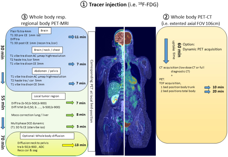
Hybrid imaging with positron emission tomography (PET) in combination with computer tomography (CT) is a well-established diagnostic tool in oncological staging and restaging. The combination of PET with magnetic resonance imaging (MRI) as a clinical scanner was introduced approximately 10 years ago. Although MRI provides superb soft tissue contrast and functional information without the radiation exposure of CT, PET-MRI is not as widely introduced in oncologic imaging as PET-CT. One reason for this hesitancy lies in the relatively long acquisition times for a PET-MRI scan, if the full diagnostic potential of MRI is exploited. In this review, we discuss the possible advantages of combined imaging protocols of PET-CT and PET-MRI, within the context of staging and restaging of patients under immunotherapy, in order to achieve "multi-hybrid imaging" in one single patient visit.

Keywords: PET-CT; PET-MRI; immunotherapy.


Conflict of interest statement

The authors declare no conflict of interest.

Figures

Figure 1
Figure 1
Components of a “multi-hybrid-imaging” oncological protocol combining PET-CT and PET-MRI with one single application of the radiopharmaceutical. The examination protocol would begin with an initial whole-body PET-CT lasting between 10 and 20 min (right side of the figure) followed by PET-MRI. PET-MRI can be performed with a fast whole-body acquisition completed within less than 30 min (upper part of the image depicting the PET-MRI protocol—blue background). To significantly take advantage of the MR-component, local PET-MRI imaging of defined body regions lasting approximately 20 min (middle part of the image depicting the PET-MRI protocol—green background) can be added. Finally, the PET-MRI examination can be further expanded with the generation of DWI of the body trunk with a minimum acquisition time of 13 min (lower part of the image depicting the PET-MRI protocol—yellow background).
Figure 2
Figure 2
Example of respiratory motion correction in PET-MRI. (A) CT: Development of a semi-solid formation post-immunotherapy at the right hilum. (B) 18F-FDG-PET with PET/CT (upper left) und PET-MRI (upper right): With MRI-based motion correction (moco), a higher SUVpeak is calculated (lower left), compared to static reconstruction (lower right).
Figure 3
Figure 3
Preferred imaging modality for the characterization of immune-related side effects (PET refers to 18F-FDG-PET).

References

    1. Beyer T., Townsend D.W., Brun T., Kinahan P.E., Charron M., Roddy R., Jerin J., Young J., Byars L., Nutt R. A combined PET/CT scanner for clinical oncology. J. Nucl. Med. 2000;41:1369–1379. - PubMed
    1. Aide N., Lasnon C., Kesner A., Levin C.S., Buvat I., Iagaru A., Hermann K., Badawi R.D., Cherry S.R., Bradley K.M., et al. New PET technologies—embracing progress and pushing the limits. Eur. J. Nucl. Med. Mol. Imaging. 2021;48:2711–2726. doi: 10.1007/s00259-021-05390-4. - DOI - PMC - PubMed
    1. Cherry S.R., Jones T., Karp J.S., Qi J., Moses W.W., Badawi R.D. Total-Body PET: Maximizing Sensitivity to Create New Opportunities for Clinical Research and Patient Care. J. Nucl. Med. 2018;59:3–12. doi: 10.2967/jnumed.116.184028. - DOI - PMC - PubMed
    1. Badawi R.D., Shi H., Hu P., Chen S., Xu T., Price P.M., Ding Y., Spencer B.A., Nardo L., Liu W., et al. First Human Imaging Studies with the EXPLORER Total-Body PET Scanner. J. Nucl. Med. 2019;60:299–303. doi: 10.2967/jnumed.119.226498. - DOI - PMC - PubMed
    1. Alberts I., Hunermund J.N., Prenosil G., Mingels C., Bohn K.P., Viscione M., Sari H., Vollnberg B., Shi K., Afshar-Oromieh A., et al. Clinical performance of long axial field of view PET/CT: A head-to-head intra-individual comparison of the Biograph Vision Quadra with the Biograph Vision PET/CT. Eur. J. Nucl. Med. Mol. Imaging. 2021;48:2395–2404. doi: 10.1007/s00259-021-05282-7. - DOI - PMC - PubMed

LinkOut - more resources