. 2021 Nov 30;13(23):6033.
doi: 10.3390/cancers13236033.

Pre-Clinical In Vitro Models Used in Cancer Research: Results of a Worldwide Survey

Affiliations

Pre-Clinical In Vitro Models Used in Cancer Research: Results of a Worldwide Survey

Sarai Martinez-Pacheco et al. Cancers (Basel). .

Abstract

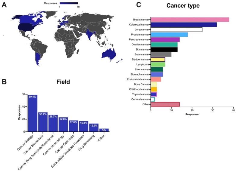
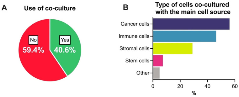
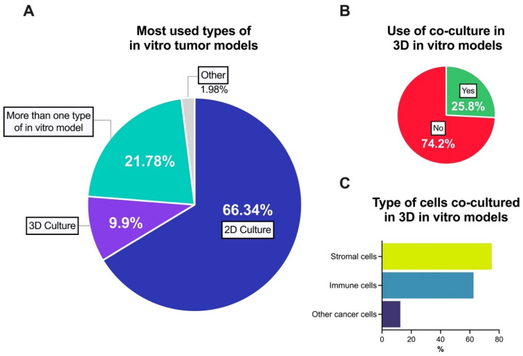
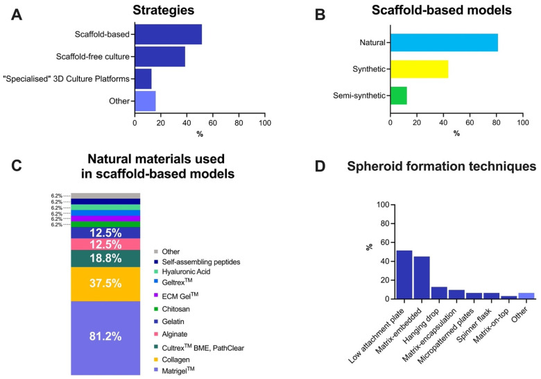
To develop and subsequently get cancer researchers to use organotypic three-dimensional (3D) models that can recapitulate the complexity of human in vivo tumors in an in vitro setting, it is important to establish what in vitro model(s) researchers are currently using and the reasons why. Thus, we developed a survey on this topic, obtained ethics approval, and circulated it throughout the world. The survey was completed by 101 researchers, across all career stages, in academia, clinical or industry settings. It included 40 questions, many with multiple options. Respondents reported on their field of cancer research; type of cancers studied; use of two-dimensional (2D)/monolayer, 2.5D and/or 3D cultures; if using co-cultures, the cell types(s) they co-culture; if using 3D cultures, whether these involve culturing the cells in a particular way to generate spheroids, or if they use additional supports/scaffolds; techniques used to analyze the 2D/2.5D/3D; and their downstream applications. Most researchers (>66%) only use 2D cultures, mainly due to lack of experience and costs. Despite most cancer researchers currently not using the 3D format, >80% recognize their importance and would like to progress to using 3D models. This suggests an urgent need to standardize reliable, robust, reproducible methods for establishing cost-effective 3D cell culture models and their subsequent characterization.

Keywords: 3D culture; 3Rs; cancer; in vitro models; methodology; precision medicine; survey.


Conflict of interest statement

The authors declare no conflict of interest.

Figures

Figure 1
Figure 1
Expansion in the numbers of peer-reviewed published articles with the term “in vitro tumor models” (A) or “3D in vitro tumor models (B) (source: Scopus; Accessed over time, most recently 17 November 2021).
Figure 2
Figure 2
General profile of the respondents. (A) Number of respondents categorized by the country of work. The graph was obtained from 101 responses; (B) Field of cancer research performed; (C) Cancer type studied by the respondents. Note: some respondents indicated that they are involved in more than one field of research and/or work on more than one cancer type.
Figure 3
Figure 3
Co-culture use in pre-clinical in vitro tumor models. (A) Use of co-culture models and (B) types of cells used in these co-culture models together with the main cell source. Results are represented as percentages (%). Note: Figure 3B represents a question with multiple choice. Some respondents indicate that their co-culture models involved more than one type of cell.
Figure 4
Figure 4
Use of 3D in vitro models in pre-clinical cancer research. (A) Main in vitro tumor models; (B) Use of co-cultured approach related to 3D in vitro tumor models; (C) Most used cell types to co-culture with cancer cells in 3D in vitro tumor models. Note: Figure 4C represents a question with multiple choice. Some respondents indicate that their co-culture models involved more than one type of cell.
Figure 5
Figure 5
Overview of 3D culture models in cancer research. (A) Main strategies used for 3D models; (B) Types of scaffold-based models used in cancer research; (C) The most used natural materials in scaffold-based models; (D) Procedures used for spheroid formation. Note: some respondents indicated that they used more than one strategy, scaffold-based model type and/or spheroid formation technique. Results are represented as percentages (%).
Figure 6
Figure 6
Analysis of pre-clinical in vitro model and their main applications. (A) Most used techniques to analyze in vitro models.; (B) Choice of cell culture models for application in the various downstream applications. Most commonly used in vitro models used for proliferation/migration/invasion assays, drug screening analysis, angiogenesis assay, cellular uptake/release assays, immune cell response assays, extracellular vesicles in vitro functional assay and gene manipulation assays. Bars represent the percentage for each option, with respondents being able to choose more than one option as appropriate. Percentages were calculated based on the reported use of each technique.
Figure 7
Figure 7
Principal reasons for not using 3D models (%).

References

    1. Breslin S., O’Driscoll L. Three-dimensional cell culture: The missing link in drug discovery. Drug Discov. Today. 2013;18:240–249. doi: 10.1016/j.drudis.2012.10.003. - DOI - PubMed
    1. Katt M.E., Placone A.L., Wong A.D., Xu Z.S., Searson P.C. In Vitro Tumor Models: Advantages, Disadvantages, Variables, and Selecting the Right Platform. Front. Bioeng. Biotechnol. 2016;4:12. doi: 10.3389/fbioe.2016.00012. - DOI - PMC - PubMed
    1. Hait W.N. Anticancer drug development: The grand challenges. Nat. Rev. Drug Discov. 2010;9:253–254. doi: 10.1038/nrd3144. - DOI - PubMed
    1. Hopkins A.L. Network pharmacology: The next paradigm in drug discovery. Nat. Chem. Biol. 2008;4:682–690. doi: 10.1038/nchembio.118. - DOI - PubMed
    1. Kola I. The State of Innovation in Drug Development. Clin. Pharmacol. Ther. 2008;83:227–230. doi: 10.1038/sj.clpt.6100479. - DOI - PubMed

LinkOut - more resources