

Lyophilised Platelet-Rich Fibrin: Physical and Biological Characterisation
- PMID: 34885714
- PMCID: PMC8658988
- DOI: 10.3390/molecules26237131
Lyophilised Platelet-Rich Fibrin: Physical and Biological Characterisation
Abstract
Background: Platelet-rich fibrin (PRF) has gained popularity in craniofacial surgery, as it provides an excellent reservoir of autologous growth factors (GFs) that are essential for bone regeneration. However, the low elastic modulus, short-term clinical application, poor storage potential and limitations in emergency therapy use restrict its more widespread clinical application. This study fabricates lyophilised PRF (Ly-PRF), evaluates its physical and biological properties, and explores its application for craniofacial tissue engineering purposes.
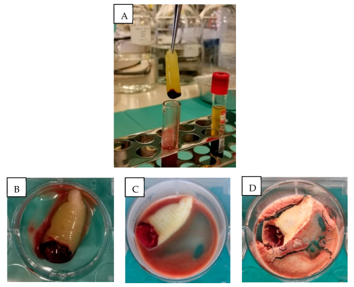
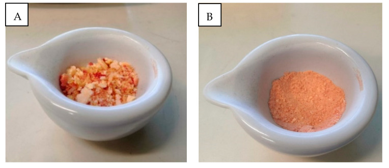
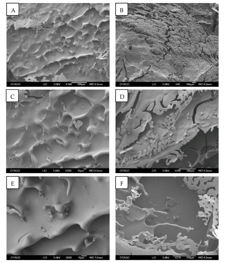
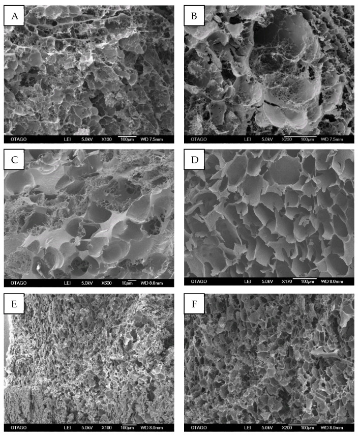
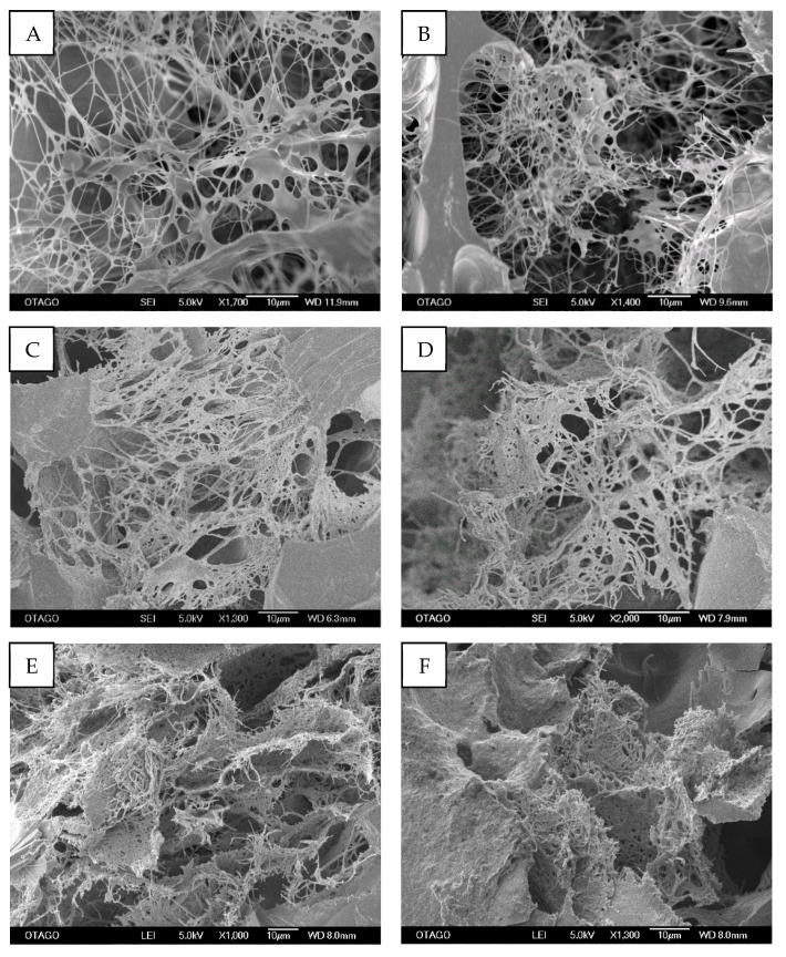
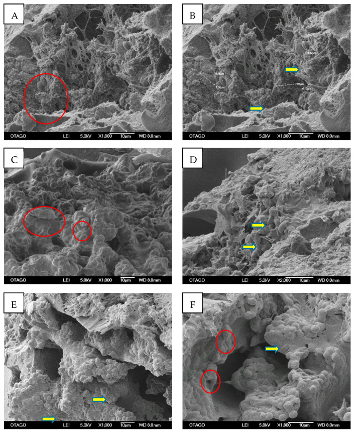
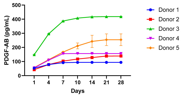
Material and methods: A lyophilisation method was applied, and the outcome was evaluated and compared with traditionally prepared PRF. We investigated how lyophilisation affected PRF's physical characteristics and biological properties by determining: (1) the physical and morphological architecture of Ly-PRF using SEM, and (2) the kinetic release of PDGF-AB using ELISA.
Results: Ly-PRF exhibited a dense and homogeneous interconnected 3D fibrin network. Moreover, clusters of morphologically consistent cells of platelets and leukocytes were apparent within Ly-PRF, along with evidence of PDGF-AB release in accordance with previously reports.
Conclusions: The protocol established in this study for Ly-PRF preparation demonstrated versatility, and provides a biomaterial with growth factor release for potential use as a craniofacial bioscaffold.
Keywords: craniofacial regeneration; lyophilisation; platelet concentrate; platelet-rich fibrin; tissue engineering.
Conflict of interest statement
The authors declare no conflict of interest. The funders had no role in the design of the study; in the collection, analyses, or interpretation of data; in the writing of the manuscript, or in the decision to publish the results.
Figures
Similar articles
-
Impact of incubation method on the release of growth factors in non-Ca2+-activated PRP, Ca2+-activated PRP, PRF and A-PRF.J Craniomaxillofac Surg. 2019 Feb;47(2):365-372. doi: 10.1016/j.jcms.2018.10.017. Epub 2018 Nov 15. J Craniomaxillofac Surg. 2019. PMID: 30578012
-
The impact of the centrifuge characteristics and centrifugation protocols on the cells, growth factors, and fibrin architecture of a leukocyte- and platelet-rich fibrin (L-PRF) clot and membrane.Platelets. 2018 Mar;29(2):171-184. doi: 10.1080/09537104.2017.1293812. Epub 2017 Apr 24. Platelets. 2018. PMID: 28437133
-
Clinical and immunohistochemical performance of lyophilized platelet-rich fibrin (Ly-PRF) on tissue regeneration.Clin Implant Dent Relat Res. 2017 Jun;19(3):466-477. doi: 10.1111/cid.12473. Epub 2017 Feb 13. Clin Implant Dent Relat Res. 2017. PMID: 28192870
-
From Blood to Regenerative Tissue: How Autologous Platelet-Rich Fibrin Can Be Combined with Other Materials to Ensure Controlled Drug and Growth Factor Release.Int J Mol Sci. 2021 Oct 26;22(21):11553. doi: 10.3390/ijms222111553. Int J Mol Sci. 2021. PMID: 34768984 Free PMC article. Review.
-
Application of Platelet Rich Fibrin in Tissue Engineering: Focus on Bone Regeneration.Platelets. 2021 Feb 17;32(2):183-188. doi: 10.1080/09537104.2020.1869710. Epub 2021 Feb 12. Platelets. 2021. PMID: 33577378 Review.
Cited by
-
Rescuing "hopeless" avulsed teeth using autologous platelet-rich fibrin following delayed reimplantation: Two case reports.World J Clin Cases. 2023 Jan 26;11(3):635-644. doi: 10.12998/wjcc.v11.i3.635. World J Clin Cases. 2023. PMID: 36793624 Free PMC article.
-
Insights Into the Role of Platelet-Derived Growth Factors: Implications for Parkinson's Disease Pathogenesis and Treatment.Front Aging Neurosci. 2022 Jul 1;14:890509. doi: 10.3389/fnagi.2022.890509. eCollection 2022. Front Aging Neurosci. 2022. PMID: 35847662 Free PMC article. Review.
-
Platelet Power: Revitalizing Endodontics With Scaffolds.Cureus. 2024 May 20;16(5):e60691. doi: 10.7759/cureus.60691. eCollection 2024 May. Cureus. 2024. PMID: 38899240 Free PMC article. Review.
-
Platelet-rich fibrin as an autologous biomaterial for bone regeneration: mechanisms, applications, optimization.Front Bioeng Biotechnol. 2024 Apr 16;12:1286035. doi: 10.3389/fbioe.2024.1286035. eCollection 2024. Front Bioeng Biotechnol. 2024. PMID: 38689760 Free PMC article. Review.
-
Effect of Currently Available Nanoparticle Synthesis Routes on Their Biocompatibility with Fibroblast Cell Lines.Molecules. 2022 Oct 17;27(20):6972. doi: 10.3390/molecules27206972. Molecules. 2022. PMID: 36296564 Free PMC article.
References
-
- Choukroun J., Diss A., Simonpieri A., Girard M.O., Schoeffler C., Dohan S.L., Anthony J.J., Jaafar M., David M. Platelet-rich fibrin (PRF): A second-generation platelet concentrate. Part IV: Clinical effects on tissue healing. Oral Surg. Oral Med. Oral Pathol. Oral Radiol. Endodontol. 2006;101:e56–e60. doi: 10.1016/j.tripleo.2005.07.011. - DOI - PubMed
-
- Dohan D.M., Choukroun J., Diss A., Dohan S.L., Dohan A.J., Mouhyi J., Gogly B. Platelet-rich fibrin (PRF): A second-generation platelet concentrate. Part II: Platelet-related biologic features. Oral Surg. Oral Med. Oral Pathol. Oral Radiol. Endodontol. 2006;101:e45–e50. doi: 10.1016/j.tripleo.2005.07.009. - DOI - PubMed
-
- Miron R.J., Choukroun J. Platelet Rich Fibrin in Regenerative Dentistry. John Wiley & Sons Ltd.; Hoboken, NJ, USA: 2017.
-
- Dohan Ehrenfest D.M., Diss A., Odin G., Doglioli P., Hippolyte M.-P., Charrier J.-B. In vitro effects of Choukroun’s PRF (platelet-rich fibrin) on human gingival fibroblasts, dermal prekeratinocytes, preadipocytes, and maxillofacial osteoblasts in primary cultures. Oral Surg. Oral Med. Oral Pathol. Oral Radiol. Endodontol. 2009;108:341–352. doi: 10.1016/j.tripleo.2009.04.020. - DOI - PubMed
MeSH terms
Substances
Grants and funding
LinkOut - more resources
Full Text Sources
Medical
