. 2021 Nov 27;26(23):7205.
doi: 10.3390/molecules26237205.

Adjusting the Structure of β-Cyclodextrin to Improve Complexation of Anthraquinone-Derived Drugs

Affiliations

Adjusting the Structure of β-Cyclodextrin to Improve Complexation of Anthraquinone-Derived Drugs

Agata Krzak et al. Molecules. .

Abstract

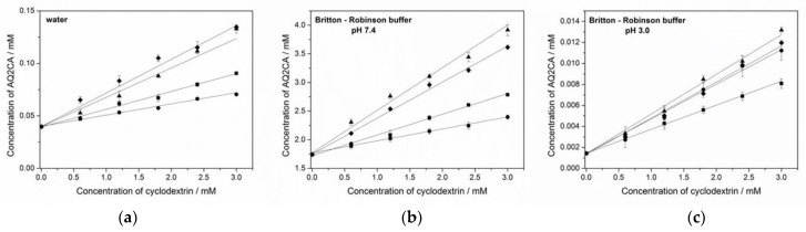
β-Cyclodextrin (CD) derivatives containing an aromatic triazole ring were studied as potential carriers of the following drugs containing an anthraquinone moiety: anthraquinone-2-sulfonic acid (AQ2S); anthraquinone-2-carboxylic acid (AQ2CA); and a common anthracycline, daunorubicin (DNR). UV-Vis and voltammetry measurements were carried out to determine the solubilities and association constants of the complexes formed, and the results revealed the unique properties of the chosen CDs as effective pH-dependent drug complexing agents. The association constants of the drug complexes with the CDs containing a triazole and lipoic acid (βCDLip) or galactosamine (βCDGAL), were significantly larger than that of the native βCD. The AQ2CA and AQ2S drugs were poorly soluble, and their solubilities increased as a result of complex formation with βCDLip and βCDGAL ligands. AQ2CA and AQ2S are negatively charged at pH 7.4. Therefore, they were less prone to form an inclusion complex with the hydrophobic CD cavity than at pH 3 (characteristic of gastric juices) when protonated. The βCDTriazole and βCDGAL ligands were found to form weaker inclusion complexes with the positively charged drug DNR at an acidic pH (pH 5.5) than in a neutral medium (pH 7.4) in which the drug dissociates to its neutral, uncharged form. This pH dependence is favorable for antitumor applications.

Keywords: anthraquinone-2-carboxylic acid; anthraquinone-2-sulfonic acid; association constant; cyclodextrins; daunorubicin; inclusion complex; solubility.


Conflict of interest statement

The authors declare no conflict of interest.

Figures

Figure 1
Figure 1
Structures of anthraquinone-2-sulfonic acid (AQ2S), anthraquinone-2-carboxylic acid (AQ2CA), and daunorubicin (DNR).
Figure 2
Figure 2
Structures of β-cyclodextrin and its derivatives used in the present work.
Figure 3
Figure 3
The UV-Vis spectra of (a) βCDGAL, (b) βCDLip, and (c) βCDTriazol at the pH range between 2.7 and 9.5. The dependences of the absorbance on the pH of the Britton–Robinson buffer are presented in the figures.
Figure 4
Figure 4
Phase solubility diagrams of the inclusion complexes of AQ2CA with βCD (■), βCDamine (●), βCDLip (♦), and βCDGAL (▲) in: (a) Water; (b) Britton–Robinson buffer at pH 7.4; (c) Britton–Robinson buffer at pH 3.0.
Figure 5
Figure 5
Cyclic voltammograms of 2.5 × 10⁻⁵ M (a) AQ2CA and (b) AQ2S in the absence (solid line) and presence of 1.08 × 10⁻³ M βCDGAL (dashed line), recorded in BR buffer at pH 3.0. All potentials are reported vs. a silver/silver chloride (Ag/AgCl) electrode. Scan rate, 100 mV·s−1.
Figure 6
Figure 6
Osa (Equation (2)) dependencies for (a) AQ2CA and (b) AQ2S in the presence of βCDGAL at pH 7.4 (■) and 3.0 (●). Reduction peak currents were recorded using cyclic voltammetry at a scan rate of 100 mV·s−1.
Figure 7
Figure 7
Osa (Equation (2)) dependencies for DNR in the presence of βCDGAL at pH 7.4 (■) and pH 5.5 (●).
Figure 8
Figure 8
1H NOESY spectrum of the βCDGAL/AQ2S system. The cross-peaks between the H3 and H5 internal protons of βCDGAL and the protons in the aromatic rings of AQ2S are marked by the blue rectangle. In this spectrum, there is a lack of evidence for interactions between the external protons of βCDGAL, i.e., H1, H2, and H4, and protons of the ligand, AQ2S (red rectangles, no signals present). The signal of HDO is marked by an asterisk.
Scheme 1
Scheme 1
Oxidation and reduction reactions of: (A) AQ2CA and AQ2S; (B) DNR.

References

    1. Greidanus J., Willemse P.H., Uges D.R., Oremus E.T., De Langen Z.J., De Vries E.G. Continuous infusion of low-dose doxorubicin, epirubicin and mitoxantrone in cancer chemotherapy: A review. Pharm. Weekbl. 1988;10:237–245. doi: 10.1007/BF02219662. - DOI - PubMed
    1. Patterson L.H. Bioreductively activated antitumor N-oxides: The case of AQ4N, a unique approach to hypoxia-activated cancer chemotherapy. Drug Metab. Rev. 2002;34:581–592. doi: 10.1081/DMR-120005659. - DOI - PubMed
    1. Andersen D.O., Weber N.D., Wood S.G., Hughes B.G., Murray B.K., North J.A. In vitro virucidal activity of selected anthraquinones and anthraquinone derivatives. Antivir. Res. 1991;16:185–196. doi: 10.1016/0166-3542(91)90024-L. - DOI - PubMed
    1. Panigrahi G.K., Yadav A., Mandal P., Tripathi A., Das M. Immunomodulatory potential of Rhein, an anthraquinone moiety of Cassia occidentalis seeds. Toxicol. Lett. 2016;245:15–23. doi: 10.1016/j.toxlet.2016.01.006. - DOI - PubMed
    1. Kshirsagar A.D., Panchal P.V., Harle U.N., Nanda R.K., Shaikh H.M. Anti-inflammatory and antiarthritic activity of anthraquinone derivatives in rodents. Int. J. Inflamm. 2014;2014:690596. doi: 10.1155/2014/690596. - DOI - PMC - PubMed