

Immunohistochemical Characterization of a Duodenal Adenocarcinoma with Pulmonary, Hepatic and Parapatellar Metastases in a Common Marmoset (Callithrixjacchus)
- PMID: 34886977
- PMCID: PMC8669625
- DOI: 10.1016/j.jcpa.2021.09.002
Immunohistochemical Characterization of a Duodenal Adenocarcinoma with Pulmonary, Hepatic and Parapatellar Metastases in a Common Marmoset (Callithrixjacchus)
Abstract
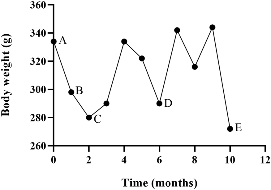
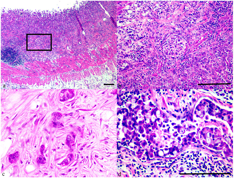
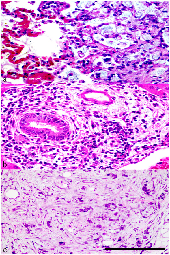
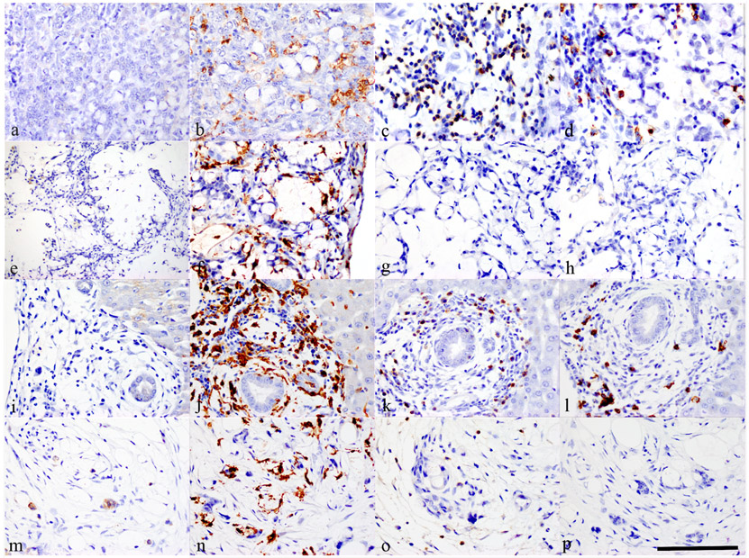
An 11-year-old male common marmoset (Callithrix jacchus) presented with chronic, progressive weight loss and diarrhoea. Response to treatment with nutritional supplementation, antibiotics and immunosuppressants was modest and transient, and the animal was humanely euthanized. At necropsy, the proximal 8 cm of small intestine was diffusely pale with transmural thickening. The lungs contained coalescing tan, firm nodules measuring up to 4 mm in diameter. Histological examination revealed infiltrative mucinous adenocarcinoma of the duodenum with extensive metastases to the lungs, liver and left parapatellar adipose tissue. The mucinous matrix secreted by the primary and metastatic lesions was strongly periodic acid-Schiff positive. Warthin Starry staining for spirochaetes was negative. Pancytokeratin expression was attenuated in the primary tumour as well as in the metastases, which correlated to a poorly differentiated phenotype. To the authors' knowledge, this is the first report of a proximal duodenal adenocarcinoma with extensive metastatic disease in a common marmoset.
Keywords: common marmoset; duodenal mucinous adenocarcinoma; metastases; pancytokeratin.
Copyright © 2021 Elsevier Ltd. All rights reserved.
Conflict of interest statement
Conflict of Interest Statement
The authors declared no potential conflicts of interest with respect to the research, authorship or publication of this article.
Figures
Similar articles
-
Extensive cutaneous metastases in a dog with duodenal adenocarcinoma.Vet Clin Pathol. 2003;32(2):88-91. doi: 10.1111/j.1939-165x.2003.tb00320.x. Vet Clin Pathol. 2003. PMID: 12833224
-
Spontaneous pulmonary adenocarcinoma in a common marmoset (Callithrix jacchus).J Med Primatol. 2021 Dec;50(6):335-338. doi: 10.1111/jmp.12540. Epub 2021 Aug 26. J Med Primatol. 2021. PMID: 34448212
-
Simultaneous Occurrence of Pancreatic Adenocarcinoma and Brunner's Gland Adenoma in a Siberian Tiger (Panthera tigris altaica).J Comp Pathol. 2015 Nov;153(4):363-7. doi: 10.1016/j.jcpa.2015.08.008. Epub 2015 Sep 28. J Comp Pathol. 2015. PMID: 26422412
-
Spontaneous mediastinal myeloid sarcoma in a common marmoset (Callithrix jacchus) and review of the veterinary literature.J Med Primatol. 2017 Apr;46(2):42-47. doi: 10.1111/jmp.12253. Epub 2017 Feb 1. J Med Primatol. 2017. PMID: 28145579 Free PMC article. Review.
-
Colonic mucinous adenocarcinoma in a tufted deer: differential diagnosis and literature review.J Vet Diagn Invest. 2022 Nov;34(6):983-989. doi: 10.1177/10406387221123007. Epub 2022 Sep 2. J Vet Diagn Invest. 2022. PMID: 36056527 Free PMC article. Review.
Cited by
-
The Common Marmoset-Biomedical Research Animal Model Applications and Common Spontaneous Diseases.Toxicol Pathol. 2022 Jul;50(5):628-637. doi: 10.1177/01926233221095449. Epub 2022 May 10. Toxicol Pathol. 2022. PMID: 35535728 Free PMC article.
References
-
- Abrahams N, Halverson A, Fazio V, Rybucki L, Goldblum J (2002) Adenocarcinoma of the small bowel. Diseases of the Colon and Rectum, 45, 1496–1502. - PubMed
-
- Brack M (1998) Gastrointestinal tumors observed in nonhuman primates at the German primate center. Journal of Medical Primatology, 27, 319–324. - PubMed
-
- Cabana F, Maguire R, Hsu C-D, Plowman A (2018) Identification of possible nutritional and stress risk factors in the development of marmoset wasting syndrome. Zoo Biology, 37, 98–106. - PubMed
Publication types
MeSH terms
Grants and funding
LinkOut - more resources
Full Text Sources
Medical
