Review
. 2022 Feb;47(2):149-159.
doi: 10.1016/j.tibs.2021.10.006. Epub 2021 Dec 7.

Structural parasitology of the malaria parasite Plasmodium falciparum

Affiliations
Review

Structural parasitology of the malaria parasite Plasmodium falciparum

Leonie Anton et al. Trends Biochem Sci. 2022 Feb.

Abstract

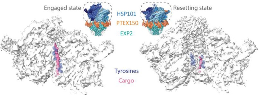
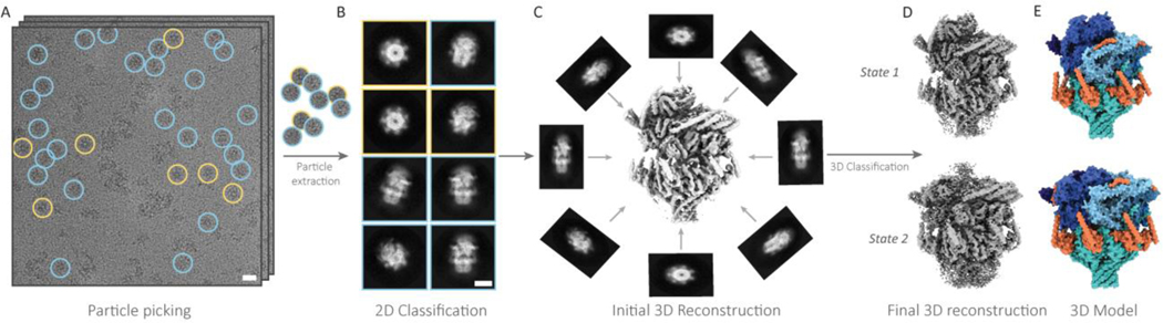
The difficulty of faithfully recapitulating malarial protein complexes in heterologous expression systems has long impeded structural study for much of the Plasmodium falciparum proteome. However, recent advances in single-particle cryo electron microscopy (cryoEM) now enable structure determination at atomic resolution with significantly reduced requirements for both sample quantity and purity. Combined with recent developments in gene editing, these advances open the door to structure determination and structural proteomics of macromolecular complexes enriched directly from P. falciparum parasites. Furthermore, the combination of cryoEM with the rapidly emerging use of in situ cryo electron tomography (cryoET) to directly visualize ultrastructures and protein complexes in the native cellular context will yield exciting new insights into the molecular machinery underpinning malaria parasite biology and pathogenesis.

Keywords: cryoEM; endogenous structure determination; in situ cryoET; malaria.


Conflict of interest statement

Declaration of interests The authors declare no conflicts of interest.

Figures

Figure 1 |
Figure 1 |. P. falciparum cryoEM protein structures from recombinant expression systems.
A, Surface representation of PfCRT cryoEM structure (PDB ID: 6UKJ, [46]). Substituted amino acids in cavity, resulting from genetic mutations that enable chloroquine (CQ) and piperaquine (PPQ) binding, are colored light blue. DV: digestive vacuole. B, CryoEM structure of PfMSP1 dimer [47]. Left protomer shows reconstruction of the six different monomer conformations found in sample, with arrows indicating flexibility (EMD-11150–55). The right protomer is shown as a cylindrical representation of the atomic model (PDB ID: 6ZBJ). Asterisk indicates interface between two protomers, which is highlighted in light pink. C, CryoEM reconstruction of Rh5-CyRPA-Ripr-Basigin complex ([48], EMD-9192). Cylindrical representation for Rh5 (light pink), CyRPA (coral), Ripr (salmon), and Basigin binding site is indicated with an arrow (PDB ID: 6MPV). Representations for all reconstructions and models were created in ChimeraX [103].
Figure 2 |
Figure 2 |. CryoEM of endogenous PTEX complex reveals native cargo.
CryoEM density maps of HSP101 from PTEX translocon in transparent white with cargo (pink) and interdigitating pore loop tyrosines (purple, stick representation) in the engaged and resetting states (EMD-8951, EMD-8952). Maps are bisected to show the endogenous cargo and pore loop tyrosines in the protein-unfolding channel of the HSP101 unfoldase. Comparing the positioning of these pore loops relative to the cargo between the two states suggests a model for the mechanisms by which HSP101 unfolds the cargo protein and threads it through the transmembrane channel of the translocon. Full PTEX complex structures are shown in the insets for context (PDB IDs: 6E10, 6E11).
Figure 3 |
Figure 3 |. Single-particle cryoEM data processing workflow.
A, cryoEM micrographs of a protein sample. Particles are identified, or “picked,” then extracted from the micrographs. B, Extracted particles are sorted into classes containing self-similar views, and then averaged to produce 2D class averages. C, Particles from high resolution 2D class averages are then used to calculate 3D reconstructions. D, 3D reconstructions are further classified and refined to yield one or more final high resolution cryoEM density maps.
Figure 4, Key Figure |
Figure 4, Key Figure |. CryoEM, FIB-SEM, and in situ cryoET workflow.
A, Tag-free sample preparation for endogenous structural proteomics. Protein complexes are enriched from malaria parasite lysates using sucrose gradient fractionation. Mass spectrometry and negative stain electron microscopy are used to identify fractions containing protein complexes of interest, which are then plunge frozen on cryoEM grids for B, single-particle cryoEM imaging in a high resolution 300kV transmission electron microscope (TEM). C, CryoEM analysis yields near-atomic resolution cryoEM density maps. CryoID is used to identify the protein(s) in the maps, enabling model building of atomic resolution structures. D, Sample preparation for endogenous CRISPR-tagged single-particle cryoEM. Affinity tags are inserted into the endogenous loci of proteins of interest in malaria parasites using CRISPR/Cas9 gene editing. Tagged proteins are affinity purified from parasite lysates, plunge frozen on cryoEM grids, and used for single-particle cryoEM imaging (B). E, CryoEM analysis yields near-atomic resolution cryoEM density maps, enabling atomic model building. F, Sample preparation for in situ cryoET. Proteins of interest are fluorescently tagged using CRISPR/Cas9 gene editing. The resulting transgenic P. falciparum parasites are grown in synchronous culture, and parasite-infected red blood cells (iRBCs) are isolated and plunge frozen directly on cryoEM grids. G, Vitrified iRBCs are then thinned in a dual-beam cryo focused ion beam scanning electron microscope (cryoFIB-SEM), yielding 100–200nm thick sections, called lamellae. Vitrified grids can be visualized in a cryo correlative light and electron microscope (cryoCLEM) to identify promising sites for cryoFIB milling. H, Tilt series are collected on lamella using a dose-symmetric tilt scheme ranging from −60° to +60° on a 300 kV TEM. I, Tilt series are aligned and reconstructed into 3D volumes called tomograms. 3D segmentation and subtomogram averaging (STA) are then used to reveal subcellular details at subnanometer resolutions. J, Integrating atomic resolution information from single-particle cryoEM with the cellular context from in situ cryoET provides further insights in to the molecular mechanisms underlying parasite biology and pathogenesis. High resolution reconstructions from cryoEM can be inserted into lower resolution subtomogram averages for context on immediate environment. Reconstructions can also be mapped back to the original 3D segmentation for cellular context.

References

    1. WHO, World malaria report 2018, 2018, p. 210.
    1. Uwimana A. et al. (2020) Emergence and clonal expansion of in vitro artemisinin-resistant Plasmodium falciparum kelch13 R561H mutant parasites in Rwanda. Nat Med. - PMC - PubMed
    1. Mathieu LC et al. (2020) Local emergence in Amazonia of Plasmodium falciparum k13 C580Y mutants associated with in vitro artemisinin resistance. Elife 9. - PMC - PubMed
    1. Conrad MD and Rosenthal PJ (2019) Antimalarial drug resistance in Africa: the calm before the storm? Lancet Infect Dis 19 (10), e338–e351. - PubMed
    1. Menard D. et al. (2018) Multidrug-resistant Plasmodium falciparum malaria in the Greater Mekong subregion. Lancet Infect Dis 18 (3), 238–239. - PubMed

Publication types

LinkOut - more resources