

18F-FDG PET/CT-Based Prognostic Survival Model After Surgery for Head and Neck Cancer
- PMID: 34887336
- PMCID: PMC9454462
- DOI: 10.2967/jnumed.121.262891
18F-FDG PET/CT-Based Prognostic Survival Model After Surgery for Head and Neck Cancer
Abstract
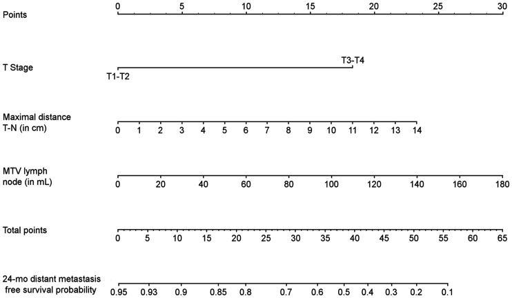
The aims of this multicenter study were to identify clinical and preoperative PET/CT parameters predicting overall survival (OS) and distant metastasis-free survival (DMFS) in a cohort of head and neck squamous cell carcinoma patients treated with surgery, to generate a prognostic model of OS and DMFS, and to validate this prognostic model with an independent cohort. Methods: A total of 382 consecutive patients with head and neck squamous cell carcinoma, divided into training (n = 318) and validation (n = 64) cohorts, were retrospectively included. The following PET/CT parameters were analyzed: clinical parameters, SUVmax, SUVmean, metabolic tumor volume (MTV), total lesion glycolysis, and distance parameters for the primary tumor and lymph nodes defined by 2 segmentation methods (relative SUVmax threshold and absolute SUV threshold). Cox analyses were performed for OS and DMFS in the training cohort. The concordance index (c-index) was used to identify highly prognostic parameters. These prognostic parameters were externally tested in the validation cohort. Results: In multivariable analysis, the significant parameters for OS were T stage and nodal MTV, with a c-index of 0.64 (P < 0.001). For DMFS, the significant parameters were T stage, nodal MTV, and maximal tumor-node distance, with a c-index of 0.76 (P < 0.001). These combinations of parameters were externally validated, with c-indices of 0.63 (P < 0.001) and 0.71 (P < 0.001) for OS and DMFS, respectively. Conclusion: The nodal MTV associated with the maximal tumor-node distance was significantly correlated with the risk of DMFS. Moreover, this parameter, in addition to clinical parameters, was associated with a higher risk of death. These prognostic factors may be used to tailor individualized treatment.
Keywords: PET/CT; distant metastasis; head and neck cancer; overall survival; prognosis.
© 2022 by the Society of Nuclear Medicine and Molecular Imaging.
Figures


References
-
- Huang SH, O’Sullivan B. Overview of the 8th edition TNM classification for head and neck cancer. Curr Treat Options Oncol. 2017;18:40. - PubMed
-
- Bray F, Ferlay J, Soerjomataram I, et al. . Global cancer statistics 2018: GLOBOCAN estimates of incidence and mortality worldwide for 36 cancers in 185 countries. CA Cancer J Clin. 2018;68:394–424. - PubMed
-
- Salaün P-Y, Abgral R, Malard O, et al. . Good clinical practice recommendations for the use of PET/CT in oncology. Eur J Nucl Med Mol Imaging. 2020;47:28–50. - PubMed
-
- Robin P, Abgral R, Valette G, et al. . Diagnostic performance of FDG PET/CT to detect subclinical HNSCC recurrence 6 months after the end of treatment. Eur J Nucl Med Mol Imaging. 2015;42:72–78. - PubMed
-
- Castelli J, Bari BD, Depeursinge A, et al. . Overview of the predictive value of quantitative 18 FDG PET in head and neck cancer treated with chemoradiotherapy. Crit Rev Oncol Hematol. 2016;108:40–51. - PubMed
Publication types
MeSH terms
Substances
LinkOut - more resources
Full Text Sources
Medical