. 2021 Dec 9;11(1):23724.
doi: 10.1038/s41598-021-02661-w.

Effect of human secretory calcium-binding phosphoprotein proline-glutamine rich 1 protein on Porphyromonas gingivalis and identification of its active portions

Affiliations

Effect of human secretory calcium-binding phosphoprotein proline-glutamine rich 1 protein on Porphyromonas gingivalis and identification of its active portions

Charline Mary et al. Sci Rep. .

Abstract

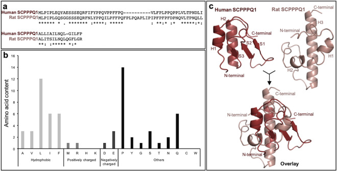
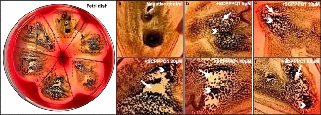
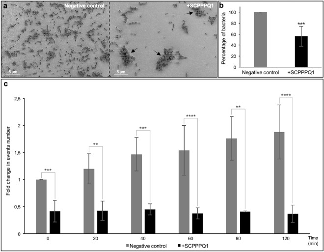
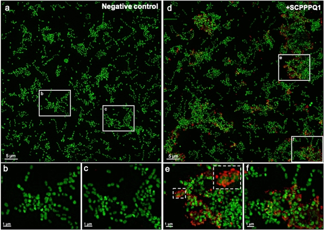
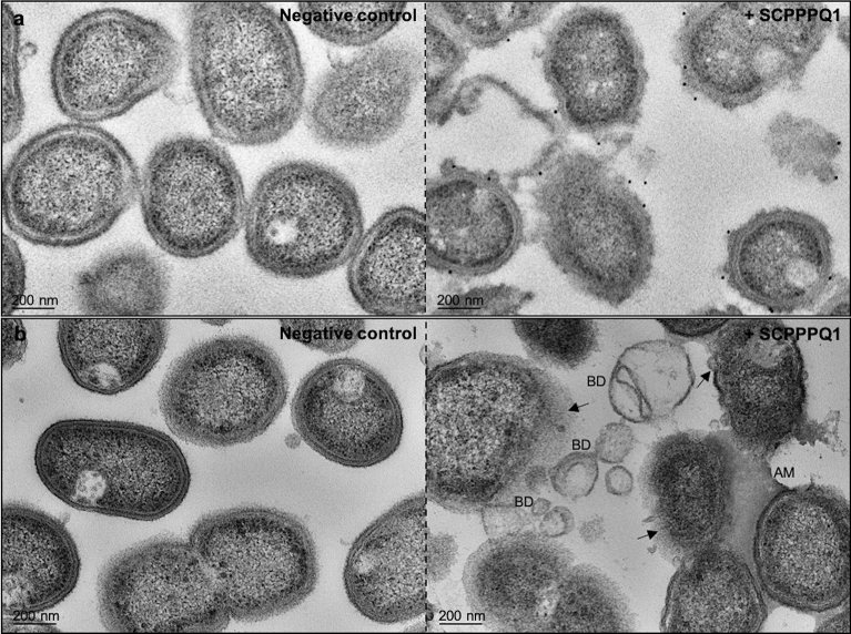
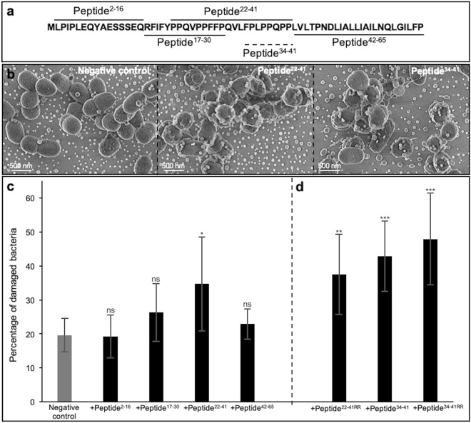
The mouth environment comprises the second most significant microbiome in the body, and its equilibrium is critical in oral health. Secretory calcium-binding phosphoprotein proline-glutamine rich 1 (SCPPPQ1), a protein normally produced by the gingival epithelium to mediate its attachment to teeth, was suggested to be bactericidal. Our aim was to further explore the antibacterial potential of human SCPPPQ1 by characterizing its mode of action and identifying its active portions. In silico analysis showed that it has molecular parallels with antimicrobial peptides. Incubation of Porphyromonas gingivalis, a major periodontopathogen, with the full-length protein resulted in decrease in bacterial number, formation of aggregates and membrane disruptions. Analysis of SCPPPQ1-derived peptides indicated that these effects are sustained by specific regions of the molecule. Altogether, these data suggest that human SCPPPQ1 exhibits antibacterial capacity and provide new insight into its mechanism of action.


Conflict of interest statement

The authors declare no competing interests.

Figures

Figure 1
Figure 1
Characterization of the human SCPPPQ1 amino acid sequence. (a) Comparison of synthetic human and rat SCPPPQ1 amino acid sequences using MUSCLE. Sequence similarity is indicated as follow: (*) identical residue (single, fully conserved), (:) conserved substitutions (strong similar properties), and (.) semi-conserved substitutions (weak similar properties). (b) In silico analysis of the human SCPPPQ1 protein using the APD3 software provided information on the distribution and relative percentage of amino acids with hydrophobic properties, positive and negative charge, and other amino acids. (c) Tridimensional structure model from i-TASSER structure prediction software for human (red) and rat (pink) SCPPPQ1 proteins and their overlay. Secondary structure elements are included (H Helix, S Sheet) and labeled according to their position in the structure (H1 to H3, S1 to S3).
Figure 2
Figure 2
Determination of the working concentration of SCPPPQ1 that affect P. gingivalis growing conditions. Comparison of the difference in growth of the bacteria on a blood agar petri dish in (a) the presence of buffer alone (negative control) or (bf) various concentrations of + SCPPPQ1. (bf) Enlargements of the boxed areas in the blood agar plate (left panel). (a) Under control conditions the bacteria formed a relatively uniform layer that appeared black in photographs, (bf) while in presence of proteins, this layer was interrupted producing a mosaic of black areas containing bacteria (white arrowhead) and white areas of various sizes that indicate absence/paucity of bacteria (white arrow). Qualitative evaluation suggests that the effect of SCPPPQ1 on the bacteria (white areas) started at 5 µM (weak) and plateaued at 20 µM (see Table 1). These results are representative of three experiments.
Figure 3
Figure 3
Effect of SCPPPQ1 on P. gingivalis bacterial population size. (a) SEM images showing bacteria from the negative control and + SCPPPQ1 treated sample at 20 min. Note the presence of aggregates only in presence of the protein (arrows). Micrographs are representative from five different experiments. (b) Quantification of the number of bacteria in percentage observed in SEM images incubated with + SCPPPQ1 normalized on the negative control at 20 min. Data are represented as mean ± standard error of mean (n = 5). Significance was determined by two-tailed Student’s t test analysis (***p < 0.001). (c) FACS results of fold change in events number over time, in samples treated with negative control or + SCPPPQ1, normalized on the negative control at 0 min. Data are represented as mean ± standard error of mean (n = 4). Significance was determined by two-tailed Student’s t test analysis (**p < 0.01; ***p < 0.001; ****p < 0.0001).
Figure 4
Figure 4
Effect of SCPPPQ1 on formation of P. gingivalis bacterial aggregates. (a) Visual inspection of bacterial cultures in test tubes from the negative control (left) and + SCPPPQ1 treated (right) samples at 20 min. A representative image from six different experiments is shown. (b) Super-resolution fluorescence images of bacteria (membrane stained in red with FM 4–64 dye) from the negative control (left) and + SCPPPQ1 treated (right) samples at 20 min. Images are representative from five different experiments. (c) FACS results of the fold change in relative aggregates volume over time normalized on the negative control at 0 min. Data are represented as mean ± standard error of mean (n = 4). Significance was determined by two-tailed Student’s t test analysis (**p < 0.01; ***p < 0.001; ****p < 0.0001).
Figure 5
Figure 5
Localisation of SCPPPQ1 on P. gingivalis using super-resolution fluorescence imaging. (ac) Images of bacteria incubated with the buffer only (negative control) at 20 min. (b,c) Enlargements of corresponding boxed areas in (a). (df) Images of bacteria incubated with + SCPPPQ1 at 20 min. (e, f) Enlargements of corresponding boxed areas in (d). (e) Upper dashed box is a magnification of the lower one. Bacterial nucleic acids were labeled with SYTO 9 (green fluorescence) and SCPPPQ1 with Alexa Fluor-546 secondary antibody (red fluorescence). These images are representative from four different experiments.
Figure 6
Figure 6
Localisation and effect of SCPPPQ1 on the surface of P. gingivalis using SEM. (a) SEM micrographs of colloidal gold immunolabeled preparation showing the presence of gold particles (white dot) on bacteria from samples treated with + SCPPPQ1 (right) and their paucity in the negative control (left) at 20 min. (b) SEM micrographs showing the normal appearance of the bacterial surface in the negative control (left) and its disruption of the bacterial surface in samples treated with + SCPPPQ1 (right) at 20 min. Micrographs are representative from four different experiments.
Figure 7
Figure 7
Localisation and effect of SCPPPQ1 on P. gingivalis bacteria using TEM imaging. (a) TEM micrographs of pre-embedded immunogold preparations of bacteria from negative control (left) and + SCPPPQ1 treated (right) samples at 20 min. No or very few gold particles (black dots) are present in the negative control while particles are associated with the bacterial surface in + SCPPPQ1 samples. (b) TEM micrographs of bacteria sections from the negative control (left) and + SCPPPQ1 treated (right) samples at 20 min. Following treatment with + SCPPPQ1, alteration of bacterial membrane (arrows), bacterial debris (BD) and aggregated material (AM) can be visualized. Micrographs are representative from three different experiments.
Figure 8
Figure 8
Effect and localisation of SCPPPQ1 on other oral bacteria. (a,c,e,g) SEM micrographs and (b,d,f,h) Super-resolution fluorescence images of (a,b) A. actinomycetemcomitans, (c,d) F. nucleatum, (e,f) P. intermedia and (g,h) T. denticola from the negative control (left) and + SCPPPQ1 treated (right) samples at 20 min. Bacterial nucleic acids were labeled with SYTO 9 (green fluorescence) and SCPPPQ1 with Alexa Fluor-546 secondary antibody (red fluorescence). Images are representative from three different experiments.
Figure 9
Figure 9
Effect of peptides on the membrane of P. gingivalis. (a) Amino acid sequence of full-length SCPPPQ1 and the corresponding five peptides generated. Peptide names are derived from the amino acid position in the full-length protein and are indicated in superscript. The dotted line represents the smallest peptide created. (b) SEM micrographs showing bacteria from the negative control,  + peptide22–42, and + peptide34–41 treated samples at 2 h. Micrographs are representative from five different experiments. (c,d) Quantification of the number bacteria with damaged membrane in percentage from the negative control and + peptides treated samples. Two peptides were synthesized in which two arginines were added on the C-terminal end; these are referred to as peptide22-41RR and peptide34-41RR. Data are represented as mean ± standard error of mean (n = 5). Significance was determined by two-tailed Student’s t test analysis (ns: p > 0.05; *p < 0.05; **p < 0.01; ***p < 0.001).

References

    1. Deo PN, Deshmukh R. Oral microbiome: Unveiling the fundamentals. J. Oral Maxillofac. Pathol. 2019;23:122–128. doi: 10.4103/jomfp.JOMFP_304_18. - DOI - PMC - PubMed
    1. Lu M, Xuan S, Wang Z. Oral microbiota: A new view of body health. Food Sci. Hum. Wellness. 2019;8:8–15. doi: 10.1016/j.fshw.2018.12.001. - DOI
    1. Kumar PS. From focal sepsis to periodontal medicine: A century of exploring the role of the oral microbiome in systemic disease. J. Physiol. 2017;595:465–476. doi: 10.1113/JP272427. - DOI - PMC - PubMed
    1. Zhang L, Liu Y, Zheng HJ, Zhang CP. The oral microbiota may have influence on oral cancer. Front. Cell Infect. Microbiol. 2019;9:476. doi: 10.3389/fcimb.2019.00476. - DOI - PMC - PubMed
    1. Lo CH, et al. Periodontal disease, tooth loss, and risk of serrated polyps and conventional adenomas. Cancer Prev. Res. (Phila) 2020;13:699–706. doi: 10.1158/1940-6207.CAPR-20-0090. - DOI - PMC - PubMed

Publication types

MeSH terms

Grants and funding

LinkOut - more resources