. 2021 Dec 9;11(1):23767.
doi: 10.1038/s41598-021-03091-4.

Peptide backbone modifications of amyloid β (1-40) impact fibrillation behavior and neuronal toxicity

Affiliations

Peptide backbone modifications of amyloid β (1-40) impact fibrillation behavior and neuronal toxicity

Benedikt Schwarze et al. Sci Rep. .

Abstract

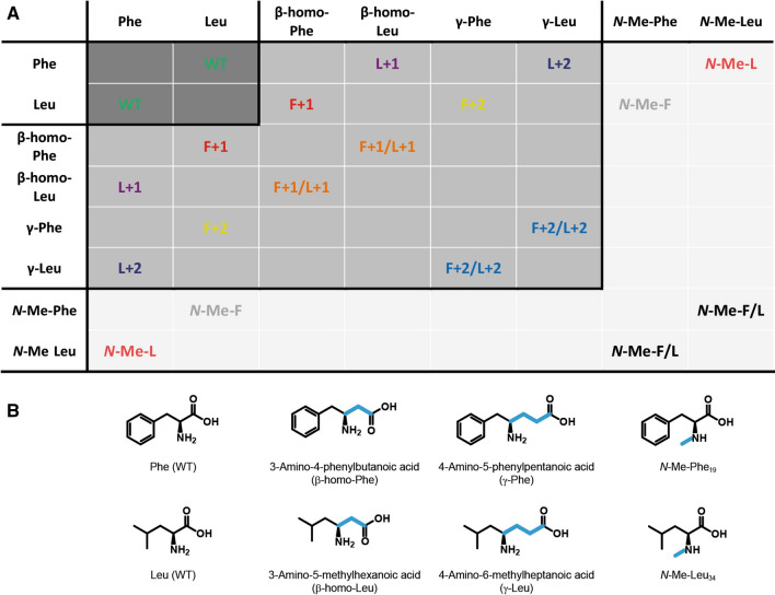
Fibril formation of amyloid β (Aβ) peptides is one of the key molecular events connected to Alzheimer's disease. The pathway of formation and mechanism of action of Aβ aggregates in biological systems is still object of very active research. To this end, systematic modifications of the Phe19-Leu34 hydrophobic contact, which has been reported in almost all structural studies of Aβ40 fibrils, helps understanding Aβ folding pathways and the underlying free energy landscape of the amyloid formation process. In our approach, a series of Aβ40 peptide variants with two types of backbone modifications, namely incorporation of (i) a methylene or an ethylene spacer group and (ii) a N-methylation at the amide functional group, of the amino acids at positions 19 or 34 was applied. These mutations are expected to challenge the inter-β-strand side chain contacts as well as intermolecular backbone β-sheet hydrogen bridges. Using a multitude of biophysical methods, it is shown that these backbone modifications lead, in most of the cases, to alterations in the fibril formation kinetics, a higher local structural heterogeneity, and a somewhat modified fibril morphology without generally impairing the fibril formation capacity of the peptides. The toxicological profile found for the variants depend on the type and extent of the modification.


Conflict of interest statement

The authors declare no competing interests.

Figures

Figure 1
Figure 1
Schematic depiction of the Aβ40 structure visualizing the backbone modifications (light blue waves) introduced and the NMR labeling schemes applied. From the N-terminus, the peptide fibrils feature an unstructured N-terminus continue with β-strand 1 which ends at the beginning of the loop region, followed by β-strand 2 and the C-terminus. Both β-strands oppose each other with the side chains of the respective amino acids interacting in a steric-zipper motif. We targeted the hydrophobic contact between Phe19 and Leu34 by introduction of backbone modifications, namely methylene or ethylene spacer or N-methylation of the amide, in both residues in different combinations (see Fig. 2). Two isotopic labeling schemes (light and dark gray spheres) were applied for the investigations of local structure and dynamics of Aβ40 fibrils by solid-state NMR.
Figure 2
Figure 2
(A) Summary of the backbone mutation scheme applied to Aβ40 peptides used in this study. The color code is used throughout the manuscript in the respective data. (B) Chemical structure of the backbone modified amino acids introduced into positions 19 and 34, respectively.
Figure 3
Figure 3
Box plot representation of the two characteristic parameters lag time (left) and fibrillation time (right) determined from ThT fluorescence fibrillation kinetics experiments. The peptide variants N-Me-F and N-Me-F/L do not fibrillate, while data for F+1/L+1 could not be fitted with the applied function (n.d. means not determined). Data from three independent experiments with three replicates each are shown. Peptide concentration was 0.125 mg mL−1 in an aqueous 25 mM sodium phosphate buffered solution (pH 7.4) containing 150 mM NaCl and 0.01%(w/v) NaN3. All normalized fluorescence intensity vs. time plots are given in the supporting information (supplementary Fig. S1). Significance was tested applying a heteroscedastic Student's t-Test using two-tailed distributions, #p ≤ 0.05; ##p ≤ 0.01; ###p ≤ 0.001 vs WT.
Figure 4
Figure 4
Box plot showing the diameters of fibrils formed from all Aβ40 peptide variants determined based on TEM images. N-Me-F and N-Me-F/L do not fibrillate (n.d. means not determined); minor second fibril population of F+1/L+1 and F+2/L+2 not shown. Significance was tested applying a heteroscedastic Student's t-Test using two-tailed distributions, #p ≤ 0.05; ##p ≤ 0.01; ###p ≤ 0.001 vs WT.
Figure 5
Figure 5
X-ray diffraction patterns of fibrils formed from the Aβ40 variants. Characteristic inter-sheet (8.3–11.2 Å) and inter-strand (4.7 Å) distances are assigned. Other reflections in the range of 2θ = 14–18° and at 21° result from salts present in the amyloid samples and are not of significance.
Figure 6
Figure 6
Overview of the 13Cα –13Cβ chemical shift differences of the labeled amino acids subdivided into the two applied labeling schemes: Val18, Phe20, Ala21, Gly33 (labeling scheme 1) and Phe19, Ala21, Ile32, Val36 (labeling scheme 2) derived from 13C–13C DARR NMR experiments. Gly33 is not shown because it is lacking a Cβ atom. The bars represent the reference values of α-helix (black), random coil (gray) and β-sheet (light gray) secondary structure as reported in the literature. Experimental data is given as varying symbols in the usual color code according to the legend. The standard error for determination of chemical shifts from 13C–13C DARR NMR experiments is ± 0.5 ppm. Other conformations, if applicable, are shown as filled forms in the respective color and shape (polymorphs of Val18 in F+1/L+1 (labeling scheme 1) are identical).
Figure 7
Figure 7
Sections from the contour plot of 13C–13C DARR NMR spectra (30 °C, MAS frequency of 11,777 Hz) of L+1 (129.6 ppm), N-Me-L (129.4 ppm) and WT (128.7 ppm) using labeling scheme 2 showing a structural change upon mutation at Leu34. Left: schematic molecular structure of Ala21 and Ile32 including labeling; center: contour plot of 13C-13C DARR NMR spectra for  +1 (black), N-Me-L (red) and WT (green); right: projection of 13C-13C DARR NMR spectra for L+1 (black), N-Me-L (red) and WT (green) along y-axis.
Figure 8
Figure 8
Box plots showing results from toxicity studies of all Aβ40 peptide variants. The MTT conversion assay (upper left) represents the proportion of metabolically active cells (alive) upon exposure to Aβ40 peptide variants; the LDH release (upper right) is indicative for plasma membrane disruption (dead cells) under the same conditions; caspase-3 staining (bottom left) is used to investigate one of the possible pathways towards cell apoptosis; neurite length (bottom right) measurements are used to examine the effect of all Aβ40 peptide variants on the outgrowth of neurites and thus the wellbeing under the applied conditions. Mutant F+1/L+1 stands out in MTT and LDH, while in the caspase-3 staining the N-methylated variants show protective effects. The neurite outgrowth behavior is not significantly impacted by any of the mutants. Significance was tested applying a heteroscedastic Student's t-Test using two-tailed distributions, *p ≤ 0.05; **p ≤ 0.01; ***p ≤ 0.001 vs control; # vs WT.

References

    1. Bartolotti N, Lazarov O. Lifestyle and Alzheimer’s disease. In: Lazarov O, Tesco G, editors. Genes, Environment and Alzheimer’s Disease. Academic Press; 2016. pp. 197–237.
    1. Doig AJ, et al. Why is research on amyloid-β failing to give new drugs for Alzheimer’s disease? ACS Chem. Neurosci. 2017;8:1435–1437. doi: 10.1021/acschemneuro.7b00188. - DOI - PubMed
    1. Busche MA, Hyman BT. Synergy between amyloid-β and tau in Alzheimer’s disease. Nat. Neurosci. 2020;23:1183–1193. doi: 10.1038/s41593-020-0687-6. - DOI - PMC - PubMed
    1. Gallardo R, Ranson NA, Radford SE. Amyloid structures: Much more than just a cross-β fold. Curr. Opin. Struct. Biol. 2020;60:7–16. doi: 10.1016/j.sbi.2019.09.001. - DOI - PubMed
    1. Lotz GP, Legleiter J. The role of amyloidogenic protein oligomerization in neurodegenerative disease. J. Mol. Med. 2013;91:653–664. doi: 10.1007/s00109-013-1025-1. - DOI - PubMed

Publication types