. 2021 Aug 24;33(16):6484-6500.
doi: 10.1021/acs.chemmater.1c01947. Epub 2021 Aug 3.

Inhibition of Amyloid Aggregation and Toxicity with Janus Iron Oxide Nanoparticles

Affiliations

Inhibition of Amyloid Aggregation and Toxicity with Janus Iron Oxide Nanoparticles

Nicholas Andrikopoulos et al. Chem Mater. .

Abstract

Amyloid aggregation is a ubiquitous form of protein misfolding underlying the pathologies of Alzheimer's disease (AD), Parkinson's disease (PD) and type 2 diabetes (T2D), three primary forms of human amyloid diseases. While much has been learned about the origin, diagnosis and management of these neurological and metabolic disorders, no cure is currently available due in part to the dynamic and heterogeneous nature of the toxic oligomers induced by amyloid aggregation. Here we synthesized beta casein-coated iron oxide nanoparticles (βCas IONPs) via a BPA-P(OEGA-b-DBM) block copolymer linker. Using a thioflavin T kinetic assay, transmission electron microscopy, Fourier transform infrared spectroscopy, discrete molecular dynamics simulations and cell viability assays, we examined the Janus characteristics and the inhibition potential of βCas IONPs against the aggregation of amyloid beta (Aβ), alpha synuclein (αS) and human islet amyloid polypeptide (IAPP) which are implicated in the pathologies of AD, PD and T2D. Incubation of zebrafish embryos with the amyloid proteins largely inhibited hatching and elicited reactive oxygen species, which were effectively rescued by the inhibitor. Furthermore, Aβ-induced damage to mouse brain was mitigated in vivo with the inhibitor. This study revealed the potential of Janus nanoparticles as a new nanomedicine against a diverse range of amyloid diseases.

Keywords: Amyloid beta; IAPP; Janus nanoparticle; alpha synuclein; iron oxide nanoparticle.


Conflict of interest statement

Competing financial interests The authors declare no conflicting financial interests.

Figures

Figure 1.
Figure 1.
Synthesis of βCas IONPs from the initially synthesized N-(2-hydroxyethyl)-2,3-dibromomaleimide. As intermediate steps, Schotten-Baumann reaction, RAFT polymerization, ligand exchange and protein adsorption reaction took place sequentially.
Figure 2.
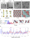
Figure 2.. Experimental and computational characterizations of βCas IONPs.
a) BN-PAGE and SDS-PAGE bands of i) BPA-P(OEGA-b-DBM) IONPs ii) βCas IONPs and iii) βCas (100 μM) b) TEM image of bare IONPs (scale bar: 50 nm) c) TEM image of βCas IONPs (scale bar: 50 nm). d) Top and side views of the BPA-P(OEGA-b-DBM) copolymer structure (backbone, yellow; PEG sidechains, pale cyan; DBM sidechains, salmon) at 5, 50, and 100 ns. e) The initial and final structures of BPA-P(OEGA-b-DBM) copolymer self-assembled monolayer (SAM). f) Radius of gyration (RG) distributions of βCas in the presence and absence of copolymer SAM. The protein is shown in cartoon. g) Averaged relative solvent accessible surface area (ΔrSASA) of βCas residues between βCas adsorbed onto the BPA-P(OEGA-b-DBM) copolymer SAM and in solution. The average number of atomic contacts (NC) between the βCas and the copolymer SAM is also plotted for comparison.
Figure 3.
Figure 3.. Inhibitory effects of βCas IONPs on Aβ, αS and IAPP fibrillization.
a, c, e) Amyloid aggregation of Aβ (50 μM), αS (50 μM) and IAPP (50 μM) in the presence and absence of βCas (25 μM) and βCas IONPs (25 μM and 12.5 μM), monitored by a ThT fluorescence kinetic assay. ThT (100 μM) was used as control and data points were depicted as mean values of repeated measurements (n=3) ± standard errors of the mean (SEM). The data points shown are the mean values (n=3) ± SEM. Statistical analysis performed through unpaired t-test determining two-tailed p-values (APA): < 0.12 (ns), < 0.033 (*), < 0.002 (**), < 0.001 (***). b, d, f) Bar graphs of t1/2, lag time, and apparent aggregation constant (k) values over time (h) for aβ, βS and IAPP in the presence and absence of βCas and βCas IONPs. Values were obtained from panels a, c & e.
Figure 4.
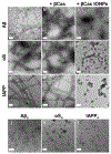
Figure 4.. TEM imaging of amyloid protein aggregation with and without βCas and βCas IONPs.
a) Aβ, b) Aβ + βCas, c) Aβ + βCas IONPs, d) αS, e) αS + βCas and f) αS + βCas IONPs, g) IAPP, h) IAPP + βCas, i) IAPP + βCas IONPs, j) oligomeric A;β (Aβo), k) oligomeric αS (αSo), l) oligomeric IAPP (IAPPo). Incubation temperature: 37 °C (scale bars: 100 nm for panels a-l). Incubation times: 21 h (a-c), 42 h (d-f), 10 h (g-i), 5 h (j), 15 h (k) and 1 h (l). Concentrations: amyloid proteins (a-i) 50 μM, βCas (a-i) 25 μM, βCas IONPs (a-i) 25 μM, and amyloid proteins (j-l) 20 μM. Amyloid protein samples (a-i) were obtained after the completion of the ThT assays presented in Figure 3a,c,e and were instantly stained on formvar/carbon-coated copper grids.
Figure 5.
Figure 5.. DMD simulations of the interactions between the three types of amyloid peptides and βCas IONPs.
For each of the three peptides – (a) Aβ, (b) IAPP, and (c) the NAC of αS, representative snapshots of the peptide encapsulated by βCas on the surface of copolymer SAM (top), the number of atomic contacts (NC) between amyloid peptide and βCas with high contact residues highlighted (middle), and the secondary structure contents of amyloid peptides with/without the presence of βCas IONP are presented.
Figure 6.
Figure 6.. Neuroblastoma and pancreatic β-cell viability compromised by exposure to Aβ, αS and IAPP, and rescued by βCas IONPs.
a, b) Significant effect of βCas IONPs (10 μM) (*** p < 0.001, ** p < 0.002) on protecting neuroblastoma cells from Aβo (20 μM; 5 h) and αSo (20 μM; 15 h) induced cell death after 20 h. Aβ (20 μM; 5 h) + βCas (10 μM) and αS (20 μM; 15 h) + βCas (10 μM) were used as negative controls. c) Prevention of IAPPo (20 μM; 1 h) cytotoxicity by βCas IONPs (10 μM) (* p < 0.033) after 15 h. IAPP (20 μM) + βCas (10 μM) was used as negative control. d) Effect of βCas IONPs (0.025-0.25 mg/mL by Fe content) on SH-SY5Y cells after 19 h. e) Effect of βCas IONPs (0.025-0.25 mg/mL by Fe content) on βTC-6 cells after 15 h. All data points are depicted as mean values (n=3) ± SEM, except for Aβ + βCas and αS + βCas, mean values (n=2) ± SEM. A statistical analysis was performed through unpaired t-test with determination of the two tailed p-values (APA). DMEM, DMEM/F-12 medium solutions with or without PBS and ultrapure H2O were used as controls.
Figure 7.
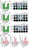
Figure 7.. Hatching survival and ROS production in embryonic zebrafish in the presence of amyloid proteins and βCas IONPs.
Mitigation of amyloid toxicity in chorion-microinjected zebrafish embryos (3 hpf) upon coadministration with βCas IONPs leading to their survival without detectable ROS production. a,c,e) Hatching survival (%) of chorionated zebrafish embryos (50 hpf) prior to direct microinjection (n=3) inside the chorion with βCas IONPs (12.5 μM), amyloid monomers Aβ, αS, IAPP) (25 μM) with and without βCas (12.5 μM) and βCas IONPs (12.5 μM). Controls were embryos treated with ThT (50 μM) or untreated (embryo medium only). b,d,f) Darkfield captures of 28 hpf and 50 hpf hatched and affected embryos, combined with images at the GFP channel of stained embryos with 1X DCFH-DA in an E3 medium solution. g,h,i) DCF fluorescence indicates significantly lower (*** p < 0.001) ROS production for zebrafish embryos microinjected with both amyloid protein and βCas IONPs, compared with the embryos exposed to amyloid protein alone. Data points are depicted as mean values (n=3) ± SEM. Statistical analysis performed through unpaired t-test determining two tailed p-values (APA): < 0.12 (ns), < 0.033 (*), < 0.002 (**), < 0.001 (***).
Figure 8.
Figure 8.. βCas IONPs alleviated Aβ aggregation and brain impairments in mice.
a) Immunofluorescence for Aβo in the hippocampus of sham-injected mice, Aβ-injected mice, Aβ-injected mice with βCas, IONP-BPA-P(OEGA-b-DBM) (IONPs-b-copolymer) or βCas IONPs. Brain slices were stained by anti-A11 antibody (green color and DAPI (blue color) was applied for nuclear counterstaining. Scale bars: 100 μm. b-d) Western blot analyses of hippocampal lysates from the brains for IL-6, IL-18, IL-1β, LC3, P62, caspase-3 expression. The gray values of the protein bands were analyzed, and quantitative data were presented as mean ± SEM.*P ≤ 0.05, **P ≤ 0.01 and ***P ≤ 0.001.

Similar articles

Cited by

References

    1. Westermark P; Sletten K; Johansson B; Cornwell GG, Fibril in senile systemic amyloidosis is derived from normal transthyretin. Proc. Natl. Acad. Sci. U S A 1990, 87, 2843–2845. - PMC - PubMed
    1. Hartl FU; Hayer-Hartl M, Converging concepts of protein folding in vitro and in vivo. Nat. Struct. Mol. Biol 2009, 16, 574–581. - PubMed
    1. Falcon B; Zivanov J; Zhang W; Murzin AG; Garringer HJ; Vidal R; Crowther RA; Newell KL; Ghetti B; Goedert M; Scheres SHW, Novel tau filament fold in chronic traumatic encephalopathy encloses hydrophobic molecules. Nature 2019, 568, 420–423. - PMC - PubMed
    1. Clark A; Lewis CE; Willis AC; Cooper GJS; Morris JF; Reid KBM; Turner RC, Islet amyloid formed from diabetes-associated peptide may be pathogenic in type-2 diabetes. The Lancet 1987, 330, 231–234. - PubMed
    1. Ke PC; Sani MA; Ding F; Kakinen A; Javed I; Separovic F; Davis TP; Mezzenga R, Implications of peptide assemblies in amyloid diseases. Chem. Soc. Rev 2017, 46, 6492–6531. - PMC - PubMed

LinkOut - more resources