. 2021 Nov 23:15:674576.
doi: 10.3389/fnins.2021.674576. eCollection 2021.

Neural Infection by Oropouche Virus in Adult Human Brain Slices Induces an Inflammatory and Toxic Response

Affiliations

Neural Infection by Oropouche Virus in Adult Human Brain Slices Induces an Inflammatory and Toxic Response

Glaucia M Almeida et al. Front Neurosci. .

Abstract

Oropouche virus (OROV) is an emerging arbovirus in South and Central Americas with high spreading potential. OROV infection has been associated with neurological complications and OROV genomic RNA has been detected in cerebrospinal fluid from patients, suggesting its neuroinvasive potential. Motivated by these findings, neurotropism and neuropathogenesis of OROV have been investigated in vivo in murine models, which do not fully recapitulate the complexity of the human brain. Here we have used slice cultures from adult human brains to investigate whether OROV is capable of infecting mature human neural cells in a context of preserved neural connections and brain cytoarchitecture. Our results demonstrate that human neural cells can be infected ex vivo by OROV and support the production of infectious viral particles. Moreover, OROV infection led to the release of the pro-inflammatory cytokine tumor necrosis factor-alpha (TNF-α) and diminished cell viability 48 h post-infection, indicating that OROV triggers an inflammatory response and tissue damage. Although OROV-positive neurons were observed, microglia were the most abundant central nervous system (CNS) cell type infected by OROV, suggesting that they play an important role in the response to CNS infection by OROV in the adult human brain. Importantly, we found no OROV-infected astrocytes. To the best of our knowledge, this is the first direct demonstration of OROV infection in human brain cells. Combined with previous data from murine models and case reports of OROV genome detection in cerebrospinal fluid from patients, our data shed light on OROV neuropathogenesis and help raising awareness about acute and possibly chronic consequences of OROV infection in the human brain.

Keywords: arboviruses; histocultures; human brain; neuroinfection; neuroinflammation; neurotropic virus; viral encephalitis.


Conflict of interest statement

The authors declare that the research was conducted in the absence of any commercial or financial relationships that could be construed as a potential conflict of interest.

Figures
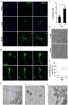
FIGURE 1
FIGURE 1
Human neural cells in adult human brain slices are susceptible to ex vivo infection by Oropouche virus (OROV). (A) Representative confocal images of uninfected control (mock) and OROV-infected slices at 24 and 48 hours post infection (hpi). Tissue sections were stained for OROV (green) and nuclei (DAPI, blue) (Scale bar = 20 μm). (B) Representative sequential Z-stack images from a cell infected by OROV in a human brain slice. Tissue was labeled for nuclei (DAPI, blue) and OROV antigen (green) (Z-Stacks = 0.5 μm) (C). Quantification of OROV positive cells at 24- and 48 hpi (n = 4–9 donors). (D) Representative cytopathic effect observed in Vero cells inoculated with the 0- or 48 hpi supernatant (103 dilution) from the one brain slice culture. (E) Virus titers in the supernatant from brain slice infected by OROV (n = 2 donors). (F) Representative transmission electron microscopy images of a human brain slice 48 hpi. Structures with typical virus particle morphology were observed (f’ and f”, highlighted by the dashed square) (Scale bar = 1 μm). Results are show as the mean ± SD. (*P < 0.05; Student t-test).
FIGURE 2
FIGURE 2
Oropouche virus infection in adult human brain slices is distributed along deep cortical layers. Adult human brain slices in culture (day in vitro 4) from middle temporal gyrus were labeled with the neuronal marker NeuN, and with anti-OROV antibody (C,D). Representative multiphoton images from an OROV-infected slice (B–D) are shown. A brightfield image from a control (non-infected) slice is also shown for reference (A). Cortical brain layers (II–VI), assigned according to neuronal morphology and density, are indicated on the left. Scale bar = 100 μm.
FIGURE 3
FIGURE 3
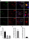
Oropouche virus targets microglia and neurons in adult human brain slices. (A) Representative confocal images of immunofluorescence labeling for OROV (green) and Iba1, NeuN or GFAP (red) in cultured slices 48 hpi. Scale bar = 20 μm. (B) Percentages of Iba1, NeuN and GFAP positive cells among total cells. (C,D) Percentage of OROV-positive cells among cells positive for Iba1 or NeuN, respectively. Results are shown as mean ± SD (n = 2–5 donors).
FIGURE 4
FIGURE 4
Differentiated human neuroblastoma cells support OROV replication. Neuroblastoma SH-SY5Y cells were differentiated into mature neurons and inoculated with OROV. (A) Cytopathic effect in SH-SY5Y cells was observed 24, 36, and 48 hpi. Images were obtained using the 20× objective. (B) Representative immunofluorescence labeling of OROV antigen (green) in SH-SY5Y cells exposed to OROV. (C) Virus titers in supernatants and monolayer at different times post infection of cultures of SH-SY5Y cell line (n = 3 biological replicates).
FIGURE 5
FIGURE 5
Oropouche virus infection induces an inflammatory and toxic response in adult human brain slices. (A) Cytokine levels in supernatants of brain slice cultures (n = 3 donors; *P < 0.05; Student t-test). (B,D) Representative immunoblots probing HLA-DR or phosphorylated Tau (pTau), respectively. Bands used for quantification are indicated with asterisks (*). β-actin was used for normalization. (C,E) HLA-DR/β-actin and pTau/β-actin ratios in four independent infected slices. Mock levels are indicated by dotted lines (n = 4 donors). (F) Cell viability in slices determined by the MTT assay 48 hpi. Actinomycin D (ActD) was used as positive control for cell death (n = 3 donors; **P < 0.001; One Way ANOVA).

References

    1. Abdullahi A. M., Sarmast S. T., Singh R. (2020). Molecular Biology and Epidemiology of Neurotropic Viruses. Cureus 12:9674. 10.7759/cureus.9674 - DOI - PMC - PubMed
    1. Acrani G. O., Gomes R., Proença-Módena J. L., da Silva A. F., Oliveira Carminati P., Silva M. L., et al. (2010). Apoptosis induced by Oropouche virus infection in HeLa cells is dependent on virus protein expression. Virus Res. 149 56–63. 10.1016/j.virusres.2009.12.013 - DOI - PubMed
    1. Al Sufiani F., Ang L. C. (2012). Neuropathology of Temporal Lobe Epilepsy. Epilep. Res. Treat. 2012:13. 10.1155/2012/624519 - DOI - PMC - PubMed
    1. Amorim M. R., Pontelli M. C., de Souza G. F., Muraro S. P., Toledo-Teixeira D. A., Forato J., et al. (2020). Oropouche virus infects, persists and induces IFN response in human peripheral blood mononuclear cells as identified by RNA PrimeFlow. And qRT-PCR assays. Viruses 12:200785. 10.3390/v12070785 - DOI - PMC - PubMed
    1. Andersson M., Avaliani N., Svensson A., Wickham J., Pinborg L. H., Jespersen B., et al. (2016). Optogenetic control of human neurons in organotypic brain cultures. Sci. Rep. 6 1–5. 10.1038/srep24818 - DOI - PMC - PubMed

LinkOut - more resources