. 2021 Nov 23:12:761418.
doi: 10.3389/fgene.2021.761418. eCollection 2021.

Wnt1 Lineage Specific Deletion of Gpr161 Results in Embryonic Midbrain Malformation and Failure of Craniofacial Skeletal Development

Affiliations

Wnt1 Lineage Specific Deletion of Gpr161 Results in Embryonic Midbrain Malformation and Failure of Craniofacial Skeletal Development

Sung-Eun Kim et al. Front Genet. .

Abstract

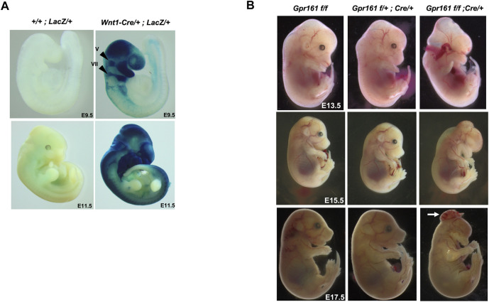
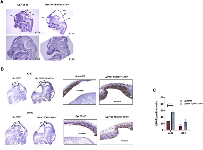
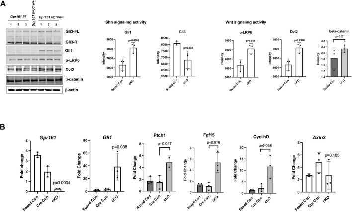
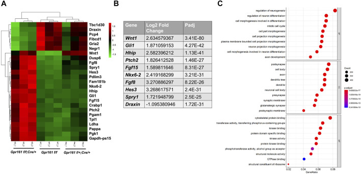
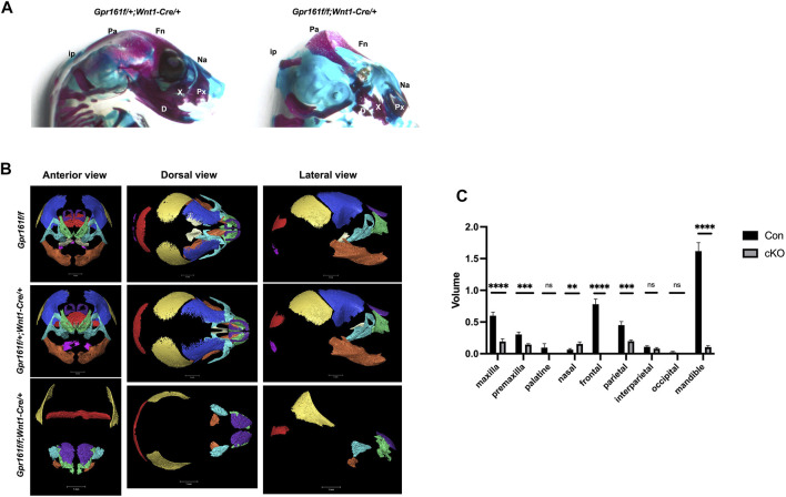
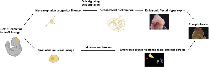
Sonic hedgehog (Shh) signaling regulates multiple morphogenetic processes during embryonic neurogenesis and craniofacial skeletal development. Gpr161 is a known negative regulator of Shh signaling. Nullizygous Gpr161 mice are embryonic lethal, presenting with structural defects involving the neural tube and the craniofacies. However, the lineage specific role of Gpr161 in later embryonic development has not been thoroughly investigated. We studied the Wnt1-Cre lineage specific role of Gpr161 during mouse embryonic development. We observed three major gross morphological phenotypes in Gpr161 cKO (Gpr161 f/f; Wnt1-Cre) fetuses; protrusive tectum defect, encephalocele, and craniofacial skeletal defect. The overall midbrain tissues were expanded and cell proliferation in ventricular zones of midbrain was increased in Gpr161 cKO fetuses, suggesting that protrusive tectal defects in Gpr161 cKO are secondary to the increased proliferation of midbrain neural progenitor cells. Shh signaling activity as well as upstream Wnt signaling activity were increased in midbrain tissues of Gpr161 cKO fetuses. RNA sequencing further suggested that genes in the Shh, Wnt, Fgf and Notch signaling pathways were differentially regulated in the midbrain of Gpr161 cKO fetuses. Finally, we determined that cranial neural crest derived craniofacial bone formation was significantly inhibited in Gpr161 cKO fetuses, which partly explains the development of encephalocele. Our results suggest that Gpr161 plays a distinct role in midbrain development and in the formation of the craniofacial skeleton during mouse embryogenesis.

Keywords: Gpr161; Wnt signaling; craniofacial defects; encephaloceles; midbrain; neural crest cells; sonic hedgehog signaling.


Conflict of interest statement

RF formerly held a leadership position in the now defunct TeratOmic Consulting LLC. He also receives travel funds from the journal Reproductive and Developmental Medicine to attend quarterly editorial board meetings. The remaining authors declare that the research was conducted in the absence of any commercial or financial relationships that could be construed as a potential conflict of interest.

Figures

FIGURE 1
FIGURE 1
The Wnt1 lineage specific deletion of Gpr161 results in protrusive tectum defects and craniofacial defects. (A) X-gal staining of Rosa-LacZ;Wnt1-Cre/+ and Rosa-LacZ at E9.5 and E11.5. V: trigeminal ganglia, VII: and facial nerve ganglia (B) Gross morphology of Gpr161 f/f , Gpr161 f/+ ;Wnt1-Cre/+, Gpr161 f/f ;Wnt1-Cre/+ (Gpr161cKO) at E13.5, 15.5 and 17.5. The white arrow indicates encephalocele in E17.5.
FIGURE 2
FIGURE 2
Histological analysis and IHC demonstrating the increased cellular proliferation in the midbrain of Gpr161 cKO fetuses. (A) H&E staining of Gpr161 f/f and Gpr161 f/f ;Wnt1-Cre/+ at E13.5 (n = 3) and E15.5 (n = 3); cb, cerebellum; cp, choroid plexus; D, mandible; HP, hard palate; mb, midbrain; NC, nasal cavity; SP, soft palate; T, tongue; tel, telecephalon; X, maxilla (B) IHC with Ki-67 and pHH3 antibodies in midbrain sections of Gpr161 f/f and Gpr161 f/f ;Wnt1-Cre/+ at E13.5 (n = 3). The black boxes indicate the areas magnified in the right panel. VZ: ventricular zone, MZ: mitotic zone (C) The statistical analysis of IHC with Ki-67 and pHH3 shown in (B). Y axis indicates the percentage of neurons that were Ki-67, pHH3, and Gli1 positive. The experiments were done triplicate and values were shown as means and standard deviations (SD). The statistical analysis was performed using a 2-way ANOVA, followed by Tukey’s test for multiple comparison (GraphPad Prism 8).
FIGURE 3
FIGURE 3
The Shh and Wnt signaling activities in the midbrain of Gpr161 cKO fetuses at E13.5. The dissected midbrain tissues from floxed control (Gpr161 f/f ), Cre control (Gpr161 f/+ ;Wnt1-Cre/+) and cKO (Gpr161 f/f ;Wnt1-Cre/+) fetuses at E13.5 were used for Western Blotting (WB) and qRT-PCR (A) Shh and Wnt signaling activities measured by WB (left). The intensity of each blot was normalized by β-actin. The quantitative analysis (n = 3) (right). Gli3 quantitation was done with repressor forms (B) qRT-PCR with Gpr161, Gli1, Ptch1, Fgf15, CyclinD, Axin2 (n = 3), which mRNA levels were normalized with Gapdh.
FIGURE 4
FIGURE 4
The transcriptomic analysis of the midbrains of Gpr161 cKO fetuses at E13.5. (A) The Heat map from dissected midbrain tissues of Gpr161 f/f (floxed control), Gpr161 f/+ ;Wnt1-Cre/+ (Cre control) and Gpr161 f/f ;Wnt1-Cre/+ (cKO) fetuses at E13.5 (n = 3). Top 25 DEGs were displayed in Heat map (Top 6 downregulated genes in Green in cKO and top 19 upregulated genes in Red in cKO). (B) Top ten differentially regulated genes (DEGs) in Gpr161 cKO (C) Gene Ontology (GO) analysis of DEGs in Gpr161 cKO.
FIGURE 5
FIGURE 5
The craniofacial skeletal analysis in Gpr161 cKO fetuses at E17.5. (A) The skeleton staining of heads from Gpr161 f/+ ;Wnt1-Cre/+ and Gpr161 f/f ;Wnt1-Cre/+ with Alcian blue and Alizarin Red S (n = 3). Fn: frontal bone; Pa: parietal bone; ip: interparietal bone; Na: nasal bone; Px: premaxilla; X: maxilla; D: mandible. (B) 3D reconstruction of microCT images of heads from Gpr161 f/f (n = 1), Gpr161 f/+ ;Wnt1-Cre/+ (n = 2) and Gpr161 f/f ;Wnt1-Cre/+ (n = 3) at E17.5. frontal bone: blue, parietal bone: yellow, interparietal bone: red, occipital bone: pink, nasal bone: purple, palatine bone: beige, maxilla: turquoise, premaxilla: green, mandible: orange (C) Quantitative volume measurement of identified craniofacial bones. Con combines the volume measurement from Gpr161 f/f and Gpr161 f/+ ;Wnt1-Cre/+.
FIGURE 6
FIGURE 6
Schematic summary of Gpr161 depletion in Wnt1-lineages in mouse. Midbrain dorsal neuroectoderm derived mesencephalon progenitor with Gpr161 depletion resulted in the increased cell proliferation via up-regulated Shh and Wnt signaling, thereby contributing to embryonic tectal protrusive phenotypes from E13.5. The Gpr161 depleted cranial neural crest cells derived from the mesencephalon caused severe cranial vault and facial bone defects. Both embryonic tectal protrusion and craniofacial bone defects could contribute to encephaloceles. This schematic figure was partly created with BioRender.com.

References

    1. Ahlgren S. C., Bronner-Fraser M. (1999). Inhibition of Sonic Hedgehog Signaling In Vivo Results in Craniofacial Neural Crest Cell Death. Curr. Biol. 9 (22), 1304–1314. 10.1016/s0960-9822(00)80052-4 - DOI - PubMed
    1. Anderson E., Peluso S., Lettice L. A., Hill R. E. (2012). Human Limb Abnormalities Caused by Disruption of Hedgehog Signaling. Trends Genet. 28 (8), 364–373. 10.1016/j.tig.2012.03.012 - DOI - PubMed
    1. Arimura N., Dewa K.-i., Okada M., Yanagawa Y., Taya S.-i., Hoshino M. (2019). Comprehensive and Cell-Type-Based Characterization of the Dorsal Midbrain during Development. Genes Cells 24 (1), 41–59. 10.1111/gtc.12656 - DOI - PubMed
    1. Bayly R. D., Ngo M., Aglyamova G. V., Agarwala S. (2007). Regulation of Ventral Midbrain Patterning by Hedgehog Signaling. Development 134 (11), 2115–2124. 10.1242/dev.02850 - DOI - PubMed
    1. Blaess S., Stephen D., Joyner A. L. (2008). Gli3 Coordinates Three-Dimensional Patterning and Growth of the Tectum and Cerebellum by Integrating Shh and Fgf8 Signaling. Development 135 (12), 2093–2103. 10.1242/dev.015990 - DOI - PMC - PubMed