. 2021 Nov 14;24(12):103460.
doi: 10.1016/j.isci.2021.103460. eCollection 2021 Dec 17.

Prolyl endopeptidase-like is a (thio)esterase involved in mitochondrial respiratory chain function

Affiliations

Prolyl endopeptidase-like is a (thio)esterase involved in mitochondrial respiratory chain function

Karen Rosier et al. iScience. .

Abstract

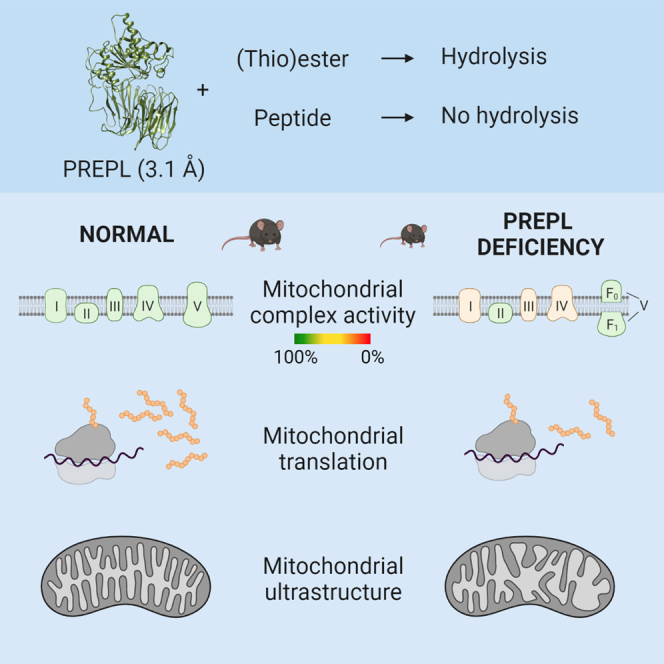
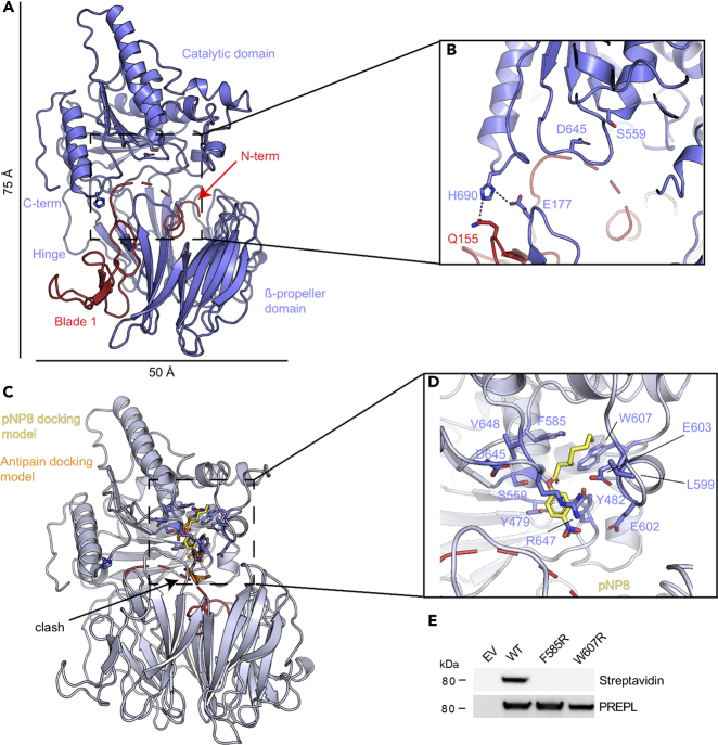
Deficiency of the serine hydrolase prolyl endopeptidase-like (PREPL) causes a recessive metabolic disorder characterized by neonatal hypotonia, feeding difficulties, and growth hormone deficiency. The pathophysiology of PREPL deficiency and the physiological substrates of PREPL remain largely unknown. In this study, we connect PREPL with mitochondrial gene expression and oxidative phosphorylation by analyzing its protein interactors. We demonstrate that the long PREPLL isoform localizes to mitochondria, whereas PREPLS remains cytosolic. Prepl KO mice showed reduced mitochondrial complex activities and disrupted mitochondrial gene expression. Furthermore, mitochondrial ultrastructure was abnormal in a PREPL-deficient patient and Prepl KO mice. In addition, we reveal that PREPL has (thio)esterase activity and inhibition of PREPL by Palmostatin M suggests a depalmitoylating function. We subsequently determined the crystal structure of PREPL, thereby providing insight into the mechanism of action. Taken together, PREPL is a (thio)esterase rather than a peptidase and PREPLL is involved in mitochondrial homeostasis.

Keywords: Molecular biology; Molecular medicine; Structural biology.


Conflict of interest statement

The authors declare no competing interests.

Figures

None
Graphical abstract
Figure 1
Figure 1
Prepl KO mice show a partial phenocopy of patients with CMS22 (A) Western blot analysis confirms the absence of PREPL expression in brain of Prepl KO mice. Intermediate expression levels are seen in heterozygous mice. (B) Weight curves of male Prepl WT and KO mice (n = 8–11) show that KO mice weigh less than WT mice from birth. (C) Prepl KO mice are smaller than WT mice. Representative example of body size difference of WT and KO male mice at 9 months. (D) Length measurement in 9-month-old male mice reveals significant differences in length of Prepl WT and KO mice (n = 7–11). (B–D) Data are shown as mean ± SEM; unpaired Student's t test; ∗∗p < 0.01; ∗∗∗p < 0.001. See also Figure S1.
Figure 2
Figure 2
PREPLL is translocated into the mitochondria (A) Thirty percent of the interaction partners of PREPL are linked to mitochondria, with interactors related to oxidative phosphorylation (OXPHOS) and mitochondrial translation. The number of PREPL protein interactors related to the large (39S) or small (28S) subunit of the mitoribosome is indicated between brackets. See also Tables S1 and S2. (B) Co-staining of endogenous PREPL (green) with Mitotracker (red) in HEK293T cells. DAPI (blue) was used for nuclear counterstaining. Endogenous PREPL is found in the cytosolic and mitochondrial fractions of HEK293T cells. An ∼2-kDa difference is observed for cytosolic and mitochondrial PREPL. Dashed line is the reference for the molecular weight of the mitochondrial form. COX4 is used as a mitochondrial marker. (C) Schematic representation of the predicted mitochondrial targeting signal (MTS, red). Predicted cleavage sites of MPP and Icp55 in PREPLL are highlighted. See also Table S3. (D) Colocalization of PREPLL (red) with MTS-GFP (green).
Figure 3
Figure 3
PREPL deficiency results in mitochondrial dysfunction (A) Activity of complexes I, III, and IV is decreased in quadriceps, cortex, and cerebellum of Prepl KO mice. Complex activities are normalized to citrate synthase (CS; n = 6–10, 12-week-old male mice). (B) RT-qPCR reveals enhanced transcription of mtDNA-encoded complex subunits in quadriceps (n = 5–6; 12-week-old male mice). β-Actin was used as a housekeeping gene. (C) Expression of CYTB and COX4 in mitochondrial fractions of Prepl KO quadriceps (n = 4). VDAC1 is used for normalization. A-C. Data are represented as mean ± SEM; Unpaired Student's t test; ∗p < 0.05, ∗∗p < 0.01, ∗∗∗p < 0.001. (D) Mitochondria in skeletal muscle of WT and Prepl KO mice. Scale bars represent 1 μm. Cristae are structured and densely packed in WT mice. Mitochondria in Prepl KO mice contain swollen or broken cristae and have more space between the cristae. Representative pictures of two WT and two KO mice. (E) Abnormal mitochondrial morphology in nerve terminal of a PREPL-deficient patient. Mitochondria appear swollen with loss of cristae and myeloid structures. Some mitochondria are engulfed in autophagosomes. The black reaction product on the junctional folds localizes AChR with peroxidase-labeled α-bungarotoxin (Régal et al., 2014). Scale bars represent 100 nm.
Figure 4
Figure 4
PREPL is inhibited by acyl protein thioesterase inhibitor Palmostatin M and has (thio)esterase activity in vitro (A) Principle of competitive ABPP. The activity-based probe FP-biotin is able to react with catalytically active PREPL. This interaction can be detected by streptavidin blotting. In the presence of an active PREPL inhibitor, FP-biotin is not able to label PREPL. (B) Palmostatin M inhibits PREPL to the same extent as inhibitor 8 (1-isobutyl-3-oxo-3,5,6,7-tetrahydro-2H-cyclopenta[c]pyridine-4-carbonitrile), a known inhibitor of PREPL. Streptavidin-labeled blot shows the interaction of PREPL with FP-biotin. The amount of GST-tagged PREPL present is determined with a GST antibody. Lanes run on same gel but are noncontiguous. Data are shown as mean ± SEM (n = 3). Differences with DMSO control were analyzed by one-way ANOVA with Dunnett's multiple comparison test. See also Figure S2. (C) PREPL cleaves the thioester substrate p-nitrophenyl thioacetate (NPTA) as efficient as APT1. Importantly, residual thioesterase activity is still observed for the PREPL S559A mutant and PREP. (D) Activity of PREPL and the inactive serine mutant PREPL S559A on p-nitrophenyl (pNP) acetate (pNP2), butyrate (pNP4), octanoate (pNP8), decanoate (pNP10), dodecanoate (pNP12), myristate (pNP14), and palmitate (pNP16). PREP is not able to hydrolyze the ester bonds. Unpaired Student's t test was performed between WT and PREPL S559A. C-D. Data are represented as the blank-corrected velocity. Data are shown as mean ± SEM (n = 3); ∗p < 0.05, ∗∗p < 0.01, ∗∗∗p < 0.001.
Figure 5
Figure 5
Crystal structure of human PREPLS contains a putative lipid-binding pocket (A) Cartoon representation of PREPLS in blue with the amino terminus and the first blade of the β-propeller (blade 1) highlighted in red. The catalytic domain containing an α/β hydrolase fold is shown on top of the β-propeller domain. The amino- and carboxy-termini are indicated, as well as the hinge region that connects the two domains. (B) The catalytic triad residues S559, D645, and H690 are found at the interface between the two domains. The displaced H690 interacts (dashed lines) with Q155 and E177 from the β-propeller domain. (C) Modeling of ligand binding into the PREPLS structure. Antipain, the ligand covalently bound to OpdB in PDB ID 4BP9, is shown as orange sticks into the equivalent position in PREPLS, where it clashes with the amino terminus of PREPLS in red. pNP8, shown as yellow sticks, is modeled bound to the proposed putative lipid-binding pocket. (D) Detailed view of the putative lipid-binding pocket, with PREPLS residues shown as blue sticks and pNP8 shown as yellow sticks. (E) The mutations of residues that form part of the putative lipid-binding pocket F585R and W607R render PREPLS inactive in the FP-biotin activity assay.

References

    1. Abrami L., Audagnotto M., Ho S., Marcaida M.J., Mesquita F.S., Anwar M.U., Sandoz P.A., Fonti G., Pojer F., Dal Peraro M., et al. Palmitoylated acyl protein thioesterase APT2 deforms membranes to extract substrate acyl chains. Nat. Chem. Biol. 2021;17:438–447. doi: 10.1038/s41589-021-00753-2. - DOI - PMC - PubMed
    1. Aertgeerts K. Crystal structure of human dipeptidyl peptidase IV in complex with a decapeptide reveals details on substrate specificity and tetrahedral intermediate formation. Protein Sci. 2004;13:412–421. doi: 10.1110/ps.03460604. - DOI - PMC - PubMed
    1. Baile M.G., Whited K., Claypool S.M. Deacylation on the matrix side of the mitochondrial inner membrane regulates cardiolipin remodeling. Mol. Biol. Cell. 2013;24:2008–2020. doi: 10.1091/mbc.E13-03-0121. - DOI - PMC - PubMed
    1. Bartholdi D., Asadollahi R., Oneda B., Schmitt-Mechelke T., Tonella P., Baumer A., Rauch A. Further delineation of genotype-phenotype correlation in homozygous 2p21 deletion syndromes: first description of patients without cystinuria. Am. J. Med. Genet. 2013;161A:1853–1859. doi: 10.1002/ajmg.a.35994. - DOI - PubMed
    1. Bartlam M., Wang G., Yang H., Gao R., Zhao X., Xie G., Cao S., Feng Y., Rao Z. Crystal structure of an acylpeptide hydrolase/esterase from aeropyrum pernix K1. Structure. 2004;12:1481–1488. doi: 10.1016/J.STR.2004.05.019. - DOI - PubMed