Case Reports
. 2022 Feb 1;163(2):bqab246.
doi: 10.1210/endocr/bqab246.

Novel Pathogenic De Novo INS p.T97P Variant Presenting With Severe Neonatal DKA

Affiliations
Case Reports

Novel Pathogenic De Novo INS p.T97P Variant Presenting With Severe Neonatal DKA

Rayhan A Lal et al. Endocrinology. .

Abstract

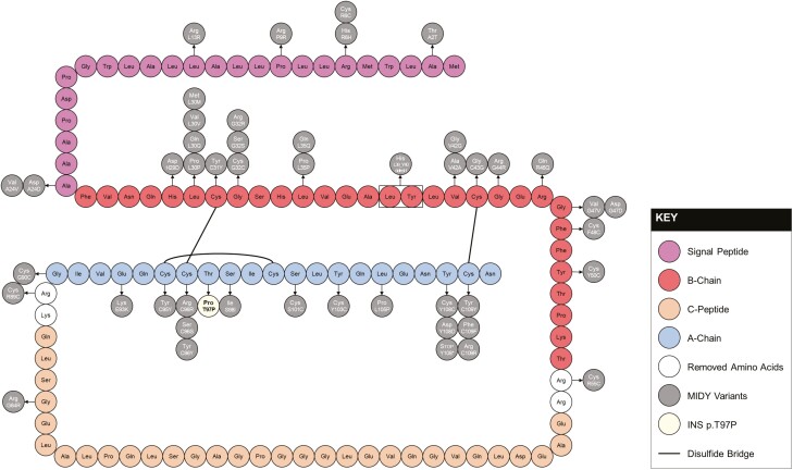
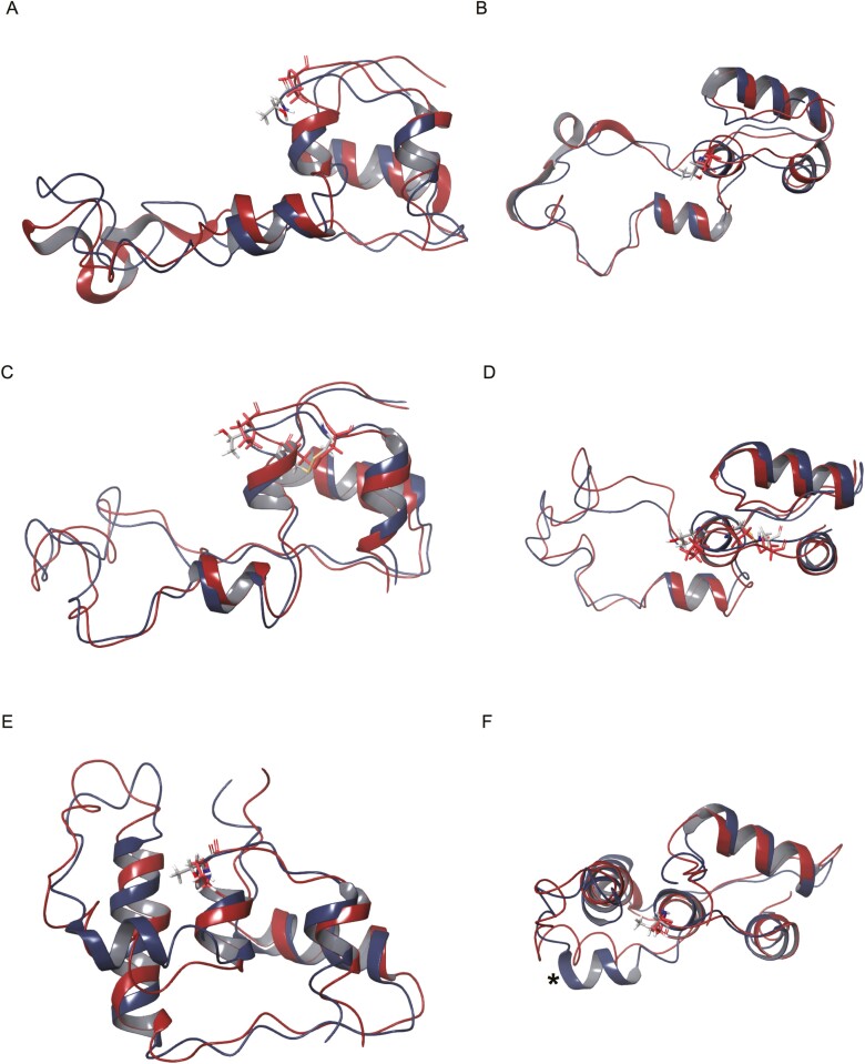
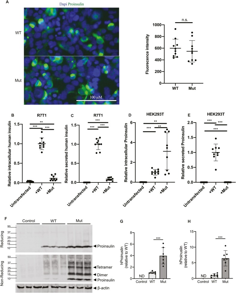
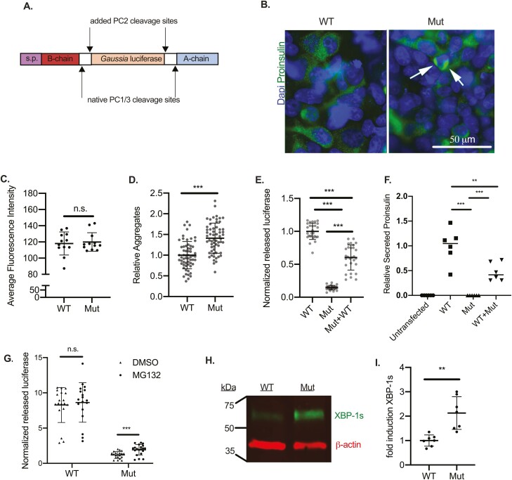
Pathogenic INS gene mutations are causative for mutant INS-gene-induced diabetes of youth (MIDY). We characterize a novel de novo heterozygous INS gene mutation (c.289A>C, p.T97P) that presented in an autoantibody-negative 5-month-old male infant with severe diabetic ketoacidosis. In silico pathogenicity prediction tools provided contradictory interpretations, while structural modeling indicated a deleterious effect on proinsulin folding. Transfection of wildtype and INS p.T97P expression and luciferase reporter constructs demonstrated elevated intracellular mutant proinsulin levels and dramatically impaired proinsulin/insulin and luciferase secretion. Notably, proteasome inhibition partially and selectively rescued INS p.T97P-derived luciferase secretion. Additionally, expression of INS p.T97P caused increased intracellular proinsulin aggregate formation and XBP-1s protein levels, consistent with induction of endoplasmic reticulum stress. We conclude that INS p.T97P is a newly identified pathogenic A-chain variant that is causative for MIDY via disruption of proinsulin folding and processing with induction of the endoplasmic reticulum stress response.

Keywords: de novo mutation; human gene mutation; insulin; insulin gene; permanent neonatal diabetes; proinsulin.


Figures

Figure 1.
Figure 1.
Schematic of preproinsulin and mutant INS-gene-induced diabetes of youth (MIDY)-causing missense mutations. Preproinsulin consists of a signal peptide (purple), B-chain (red), C-peptide (orange), and A-chain (blue). Missense variants causing MIDY are indicated with arrows and INS p.T97P is highlighted in yellow.
Figure 2.
Figure 2.
Molecular modeling of INS p.T97P proinsulin. (A-B) Lowest-energy conformer from an nuclear magnetic resonance solution of wildtype (blue) proinsulin (PDB 2KQP) compared to in silico T97P mutated proinsulin (red); (A) side view and (B) edge-on view. (C-D) Comparative homology modeling of wildtype (blue) and T97P (red) proinsulin with the Robetta structure prediction tool using PDB 2KQP as a template; (C) side view and (D) edge-on view. (E-F) Representative images of 5 models generated by the RosETTAFold de novo folding prediction tool for wildtype (blue) and T97P (red) proinsulin; (E) side view and (F) edge-on view. *Alpha helix from A81 to L86 predicted in wildtype models.
Figure 3.
Figure 3.
The INS p.T97P mutant exhibits impaired proinsulin processing and secretion. (A) Proinsulin immunostaining (left) and quantification (right) of wildtype (WT) and INS p.T97P (Mut) transfected HEK293T cells (P > 0.05, n = 3 independent experiments). (B) Relative intracellular insulin content of untransfected, WT-, or INS p.T97P-transfected R7T1 cells measured by human-specific enzyme-linked immunosorbent assay (ELISA; normalized to total cellular protein; n = 3 independent experiments). (C) Relative secreted insulin from untransfected, WT-, or INS p.T97P-transfected R7T1 cells measured by human-specific ELISA (normalized to total cellular protein; n = 3 independent experiments). (D) Relative intracellular proinsulin content of untransfected, WT-, or INS p.T97P-transfected HEK293T cells measured by proinsulin-specific ELISA (normalized to total cellular protein; n = 3 independent experiments). (E) Relative secreted proinsulin of untransfected, WT-, or INS p.T97P-transfected HEK293T cells measured by proinsulin-specific ELISA (normalized to total cellular protein; n = 3 independent experiments). (F) Representative reducing (top) and nonreducing (middle) immunoblots of proinsulin (predicted 9.4 kDa) in mock (Control), WT-, and INS p.T97P (Mut)-transfected HEK293T cells. Loading normalized to β-actin (bottom). (G, H) Quantification of (G) proinsulin under reducing conditions and (H) total reactivity under nonreducing conditions; data are normalized to β-actin (n = 3 independent experiments). Data are expressed as means, and error bars are SDs. *P ≤ 0.05; **P < 0.01; ***P ≤ 0.001, unpaired t-tests.
Figure 4.
Figure 4.
Impaired INS p.T97P processing causes endoplasmic-reticulum-associated protein degradation activation and β-cell endoplasmic reticulum stress induction. (A) Schematic of the proinsulin-luciferase reporter construct. (B) Proinsulin immunostaining of HEK293T cells transfected with the wildtype (WT) or INS p.T97P (Mut) reporter construct and (C) quantification of intracellular immunofluorescence (not significant, P =0.73). White arrows indicate proinsulin-luciferase aggregates, quantified in (D) (n = 3 independent experiments). (E) Dual luciferase assay with transfected R7T1 cells. Extracellular luciferase-insulin activity normalized to lysate CMV-firefly activity. To combine independent experiments, values were first normalized to the average dimethylsulfoxide (DMSO) value for a given experiment (n = 3 independent experiments). (F) Relative secreted proinsulin content of untransfected, WT-transfected, INSp.T97P-transfected, or WT + INSp.T97p-transfected HEK293T cells measured by proinsulin-specific enzyme-linked immunosorbent assay (normalized to total cellular protein). (G) Activity of secreted luciferase-insulin in extracellular media normalized to CMV-firefly expression in cell lysates after treatment with DMSO or MG-132 (n = 3 independent experiments). (H) Representative image of XBP-1s Western blot of HEK293T cells transfected with WT or INS p.T97P (Mut) constructs. (I) Quantification of 7 transfections (XBP-1s/actin, n = 3 independent experiments). Data are expressed as means, and error bars are SDs. *P ≤ 0.05; **P < 0.01; ***P ≤ 0.001, unpaired t-tests.

References

    1. Aguilar-Bryan L, Bryan J. Neonatal diabetes mellitus. Endocr Rev. 2008;29(3):265-291. - PMC - PubMed
    1. Dahl A, Kumar S. Recent advances in neonatal diabetes. Diabetes Metab Syndr Obes. 2020;13:355-364. - PMC - PubMed
    1. Johnson SR, McGown I, Oppermann U, Conwell LS, Harris M, Duncan EL. A novel INS mutation in a family with maturity-onset diabetes of the young: variable insulin secretion and putative mechanisms. Pediatr Diabetes. 2018;19(5):905-909. - PubMed
    1. Liu M, Hodish I, Haataja L, et al. . Proinsulin misfolding and diabetes: mutant INS gene-induced diabetes of youth. Trends Endocrinol Metab. 2010;21(11):652-659. - PMC - PubMed
    1. Liu M, Huang Y, Xu X, et al. . Normal and defective pathways in biogenesis and maintenance of the insulin storage pool. J Clin Invest. 2021;131(2):e142240. - PMC - PubMed

Publication types