. 2022 May 19;31(10):1694-1704.
doi: 10.1093/hmg/ddab352.

Canine DUXC: implications for DUX4 retrotransposition and preclinical models of FSHD

Affiliations

Canine DUXC: implications for DUX4 retrotransposition and preclinical models of FSHD

Chao-Jen Wong et al. Hum Mol Genet. .

Abstract

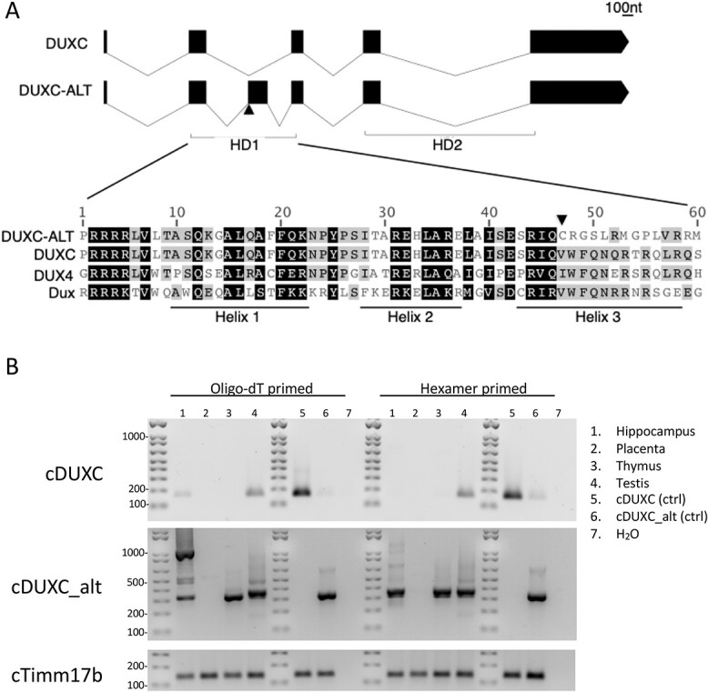
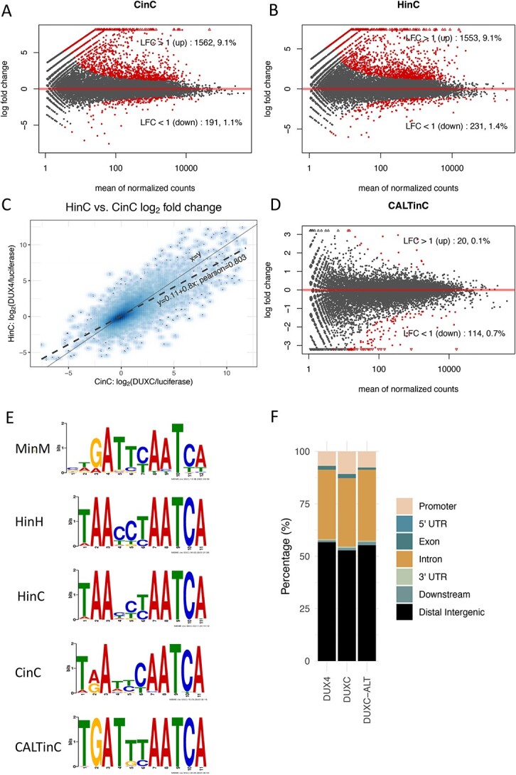
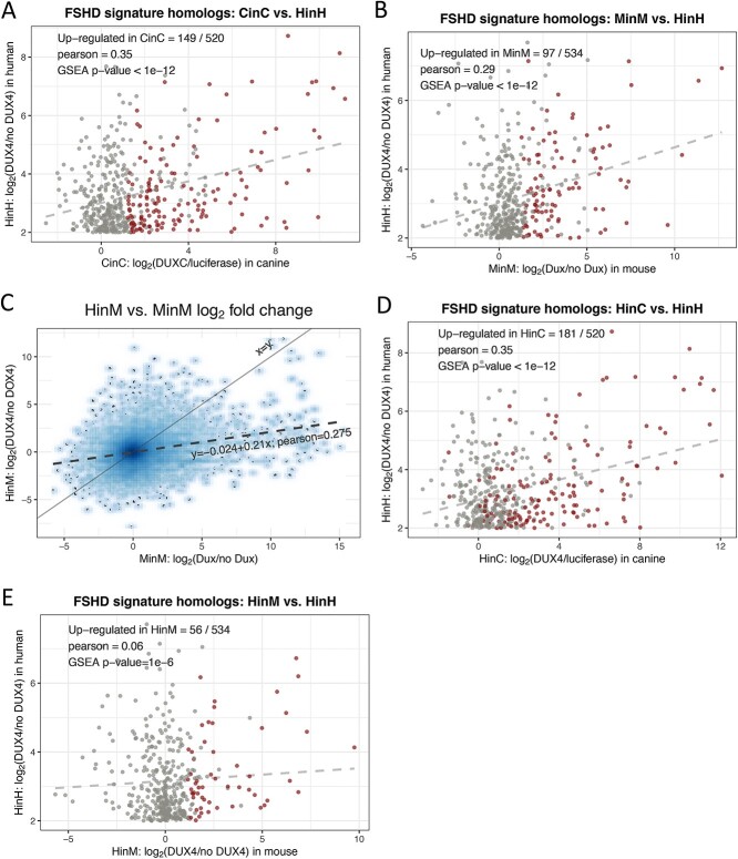
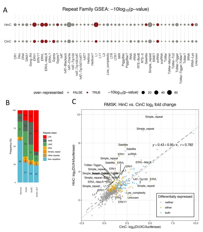
Mis-expression of DUX4 in skeletal muscle causes facioscapulohumeral muscular dystrophy (FSHD). Human DUX4 and mouse Dux are retrogenes derived from retrotransposition of the mRNA from the parental DUXC gene. Primates and rodents have lost the parental DUXC gene, and it is unknown whether DUXC had a similar role in driving an early pluripotent transcriptional program. Dogs and other Laurasiatherians have retained DUXC, providing an opportunity to determine the functional similarity to the retrotransposed DUX4 and Dux. Here, we identify the expression of two isoforms of DUXC mRNA in canine testis tissues: one encoding the canonical double homeodomain protein (DUXC), similar to DUX4/Dux, and a second that includes an in-frame alternative exon that disrupts the conserved amino acid sequence of the first homeodomain (DUXC-ALT). The expression of DUXC in canine cells induces a pluripotent program similar to DUX4 and Dux and induces the expression of a similar set of retrotransposons of the ERV/MaLR and LINE-1 families, as well as pericentromeric satellite repeats; whereas DUXC-ALT did not robustly activate gene expression in these assays. Important for preclinical models of FSHD, human DUX4 and canine DUXC show higher conservation of their homeodomains and corresponding binding motifs compared with the conservation between human DUX4 and mouse Dux, and human DUX4 activates a highly similar transcriptional program in canine cells. Together, these findings show that retrotransposition resulted in the loss of an alternatively spliced isoform and that DUXC containing mammals might be good candidates for certain preclinical models ofFSHD.


Figures

Figure 1
Figure 1
Canine DUXC locus makes two different isoforms. (A) Top panel shows a schematic of the exons encoding DUXC and DUXC-ALT and the bottom panel shows the alignment in the first homeodomain region with mouse Dux and Human DUX4 HD1. (B) Nested PCR primers specific to the DUXC (top panel) or DUXC-ALT (middle panel) mRNA isoforms show DUXC expression in testis a variably in hippocampus; DUXC-ALT is identified in testis, thymus and hippocampus.
Figure 2
Figure 2
Transcriptome and DNA binding sites of DUXC, DUX4 and DUXC-ALT in canine skeletal muscle cells. (A) A MA plot of gene expression in canine muscle cells expressing DUXC (CinC) compared with the control luciferase expression vector. The x axis is the mean of normalized counts on each gene between two conditions (A value), and the y axis is the formula image fold change (M value). Red dots represent the differentially expressed genes with adjusted P-values < 0.05 corresponding to the hypothesis formula image. (B) Similar to (A) but with comparison of the cells expressing DUX4 (HinC). (C) A smoothed scatter plot to show the linear relationship of the formula image fold change between the CinC (x axis) and HinC (y axis) comparison models. The Pearson correlation between the two sets of formula image fold change is 0.803. The gray dashed line is the linear regression, formula image, and the solid gray line is formula image. (D) Similar to (A) but with the comparison of the cells expressing DUXC-ALT (CALTinC). (E) De novo motif discovery determined binding motifs for mouse Dux expressed in mouse myoblasts (MinM), human DUX4 expressed in human myoblasts (HinH), human DUX4 expressed in canine myoblasts (HinC), and canine DUXC and DUXC-ALT expressed in canine myoblasts (CinC and CALTinC). The HinH and MinM binding motifs were derived from published ChIP-seq data GSE33838 and GSE87279, respectively. (F) Genomic distribution of ChIP-seq peaks for DUX4, DUXC and DUXC-ALT.
Figure 3
Figure 3
Comparisons to the FSHD transcriptome. (A) A scatter plot of formula image fold change of the 520 human-canine homologs of FSHD signature genes in the HinH and CinC transcriptome. Red dots indicate that the homologs are also upregulated in CinC. (B) Same as (A), but with the 534 human-mouse homologs in the HinH and MinM transcriptome, red dots indicate upregulation in MinM. (C) A smooth scatter plot to show the relationship of formula image fold change between the HinH and MinM transcriptome. The Pearson correlation between these two sets is 0.275. Gray dashed line presents the linear regression line, formula image and the solid is formula image (D) Same as (A), but with 520 human-canine homologs in the cross-species HinC and HinH transcription, and red dots indicate upregulation in HinC. (E) Same as (A), but with 534 human-mouse homologs in the HinH and cross-species HinM transcriptome, and red dots indicate upregulation inHinM.
Figure 4
Figure 4
Repeat element transcriptome shows retrotransposon activation by DUXC in canine skeletal muscle cells. (A) Repeat family enrichment analysis for DUX4 (HinC) and DUXC (CinC) expressed in canine skeletal muscle cells. Each dot represents the GSEA statistics for each repeat family with size reflecting the scaled P-value, −10log2 (P-value). Red dots indicate the family is over-represented with P-values < 0.1 and the number of significant genes in the family is higher than expected. (B) The distribution of peaks of DUX4, DUXC, DUXC-ALT transcription factors in classes of repeat elements versus the distribution of the repeat elements in the whole genome. (C) A scatter plot to show the relationship of formula image fold change of the HinC and CinC transcriptome. Each dot represents a repeat element, and the color coding presents the differential expression status. Dashed line is the linear regression line formula image, whereas the solid is formula image. A repeat element’s family name is labeled if the difference between two models is >1.5.

References

    1. Leidenroth, A., Clapp, J., Mitchell, L.M., Coneyworth, D., Dearden, F.L., Iannuzzi, L. and Hewitt, J.E. (2012) Evolution of DUX gene macrosatellites in placental mammals. Chromosoma, 121, 489–497. - PubMed
    1. Leidenroth, A. and Hewitt, J.E. (2010) A family history of DUX4: phylogenetic analysis of DUXA, B, C and Duxbl reveals the ancestral DUX gene. BMC Evol. Biol., 10, 364. - PMC - PubMed
    1. Parker, H.G., VonHoldt, B.M., Quignon, P., Margulies, E.H., Shao, S., Mosher, D.S., Spady, T.C., Elkahloun, A., Cargill, M., Jones, P.G. et al. (2009) An expressed fgf4 retrogene is associated with breed-defining chondrodysplasia in domestic dogs. Science, 325, 995–998. - PMC - PubMed
    1. Wang, C., Wallerman, O., Arendt, M.L., Sundstrom, E., Karlsson, A., Nordin, J., Makelainen, S., Pielberg, G.R., Hanson, J., Ohlsson, A. et al. (2021) A novel canine reference genome resolves genomic architecture and uncovers transcript complexity. Commun Biol, 4, 185. - PMC - PubMed
    1. Wallace, L.M., Garwick, S.E., Mei, W., Belayew, A., Coppee, F., Ladner, K.J., Guttridge, D., Yang, J. and Harper, S.Q. (2011) DUX4, a candidate gene for facioscapulohumeral muscular dystrophy, causes p53-dependent myopathy in vivo. Ann. Neurol., 69, 540–552. - PMC - PubMed

Publication types