. 2021:26:21-53.
doi: 10.1007/978-3-030-77375-5_2.

Astrocytes: The Housekeepers and Guardians of the CNS

Affiliations

Astrocytes: The Housekeepers and Guardians of the CNS

Alexei Verkhratsky et al. Adv Neurobiol. 2021.

Abstract

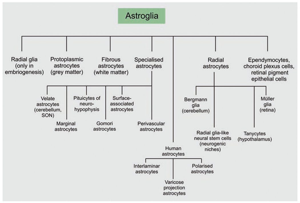
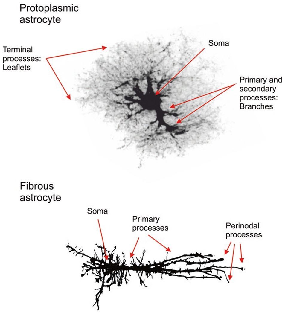
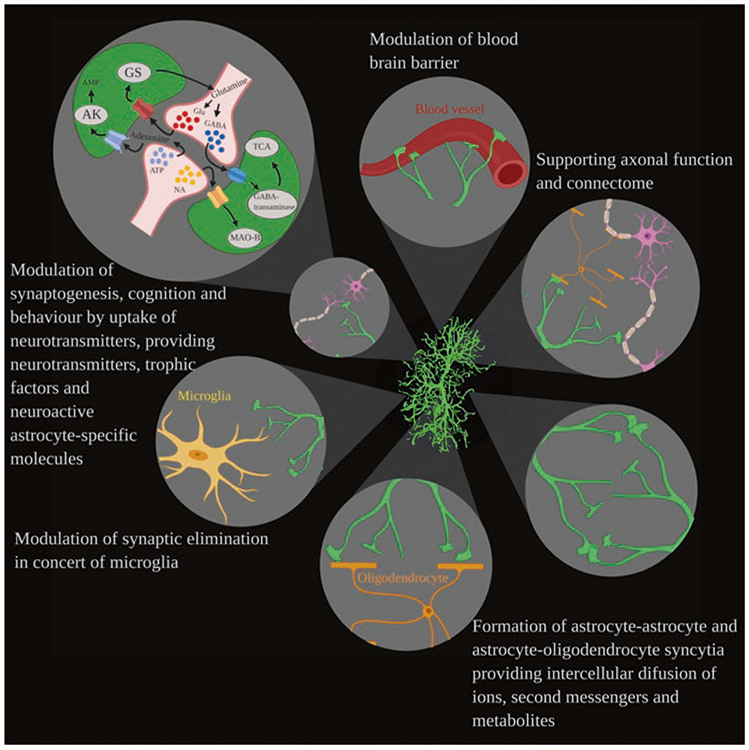
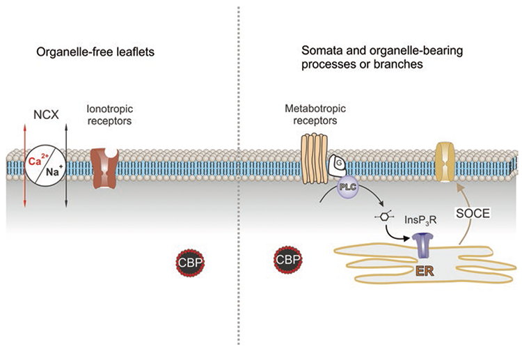
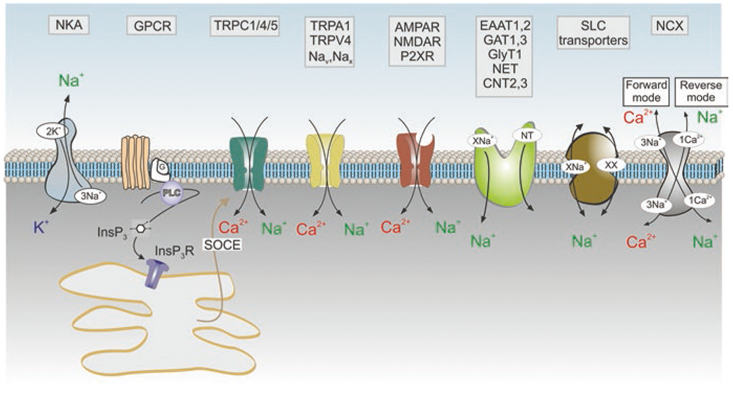
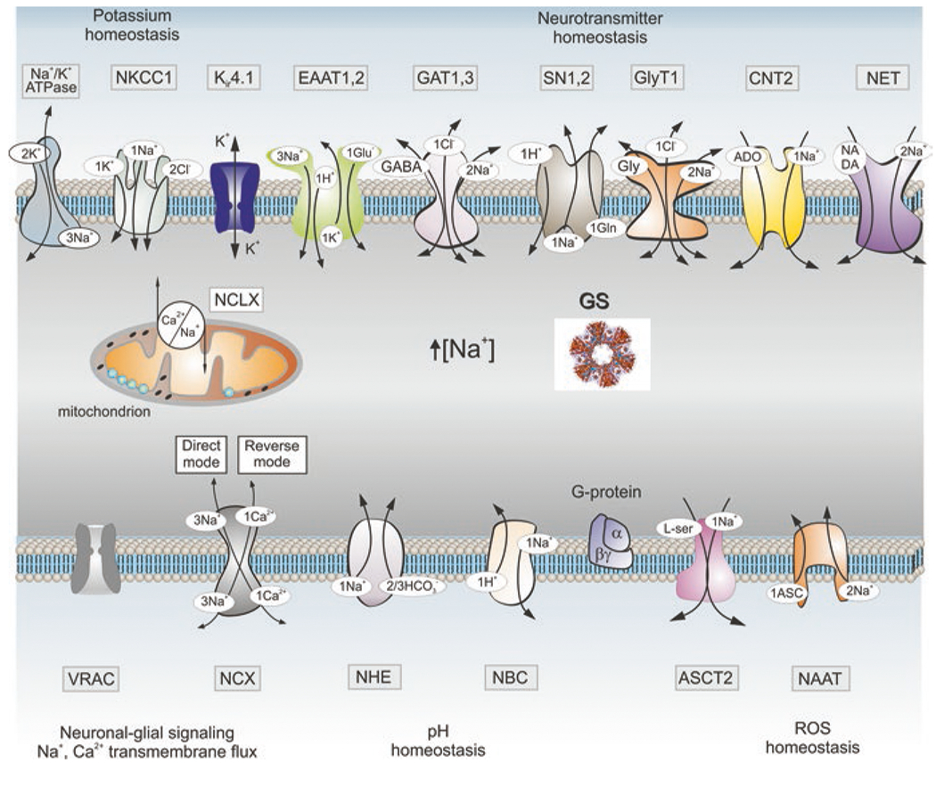
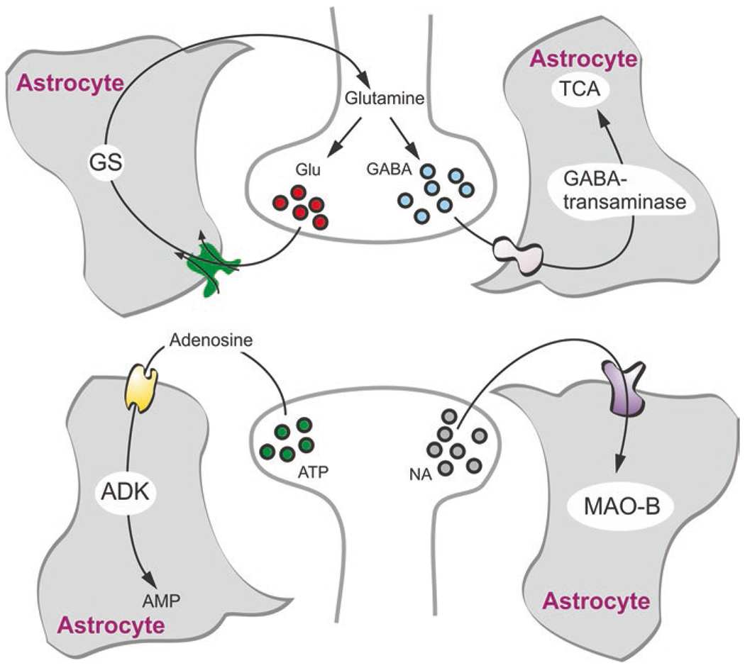
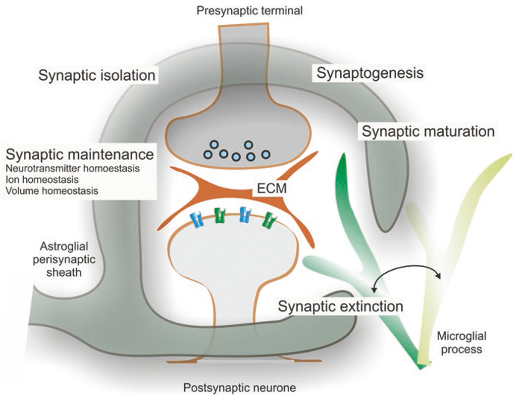
Astroglia are a diverse group of cells in the central nervous system. They are of the ectodermal, neuroepithelial origin and vary in morphology and function, yet, they can be collectively defined as cells having principle function to maintain homeostasis of the central nervous system at all levels of organisation, including homeostasis of ions, pH and neurotransmitters; supplying neurones with metabolic substrates; supporting oligodendrocytes and axons; regulating synaptogenesis, neurogenesis, and formation and maintenance of the blood-brain barrier; contributing to operation of the glymphatic system; and regulation of systemic homeostasis being central chemosensors for oxygen, CO2 and Na+. Their basic physiological features show a lack of electrical excitability (inapt to produce action potentials), but display instead a rather active excitability based on variations in cytosolic concentrations of Ca2+ and Na+. It is expression of neurotransmitter receptors, pumps and transporters at their plasmalemma, along with transports on the endoplasmic reticulum and mitochondria that exquisitely regulate the cytosolic levels of these ions, the fluctuation of which underlies most, if not all, astroglial homeostatic functions.

Keywords: Astroglia; Brain homoeostasis; Ca2+ signalling; Ion channels; Na+ signalling; Neurotransmitter receptors; SLC transporters.


Figures

Fig. 1
Fig. 1
Classification of astroglia
Fig. 2
Fig. 2
Protoplasmic and fibrous astrocytes. Protoplasmic astrocytes of the grey matter are composed of soma, primary processes or branches and organelle-free thin leaflets. Fibrous astrocytes of the white matter contain perinodal processes by which they contact oligodendrocytes and nodes of Ranvier. The protoplasmic astrocyte was traced using image provided by Milos Pekny (Gothenburg University); the camera lucida image for fibrous astrocyte was provided by Arthur Butt from University of Portsmouth
Fig. 3
Fig. 3
Functions of astrocytes. Astrocytes interact with diverse cellular structures including blood vessels, oligodendrocytes, neurones, microglia and other astrocytes, contributing to various functions (clockwise from the top) such as regulation of the blood-brain barrier, supporting axons, myelination and connectome, forming astrocyte-astrocyte and astrocyte-oligodendrocyte syncytia, controlling (in concert with microglia) synaptic elimination and modulating synaptogenesis, cognition and behaviour by the neurochemical dialogue with synapses. (Reproduced from Augusto-Oliveira et al. (2020))
Fig. 4
Fig. 4
Passive membrane properties of astrocytes. (a) Voltage-clamp recordings from astrocytes freshly isolated from the cortex of transgenic mice expressing EGFP under control of the GFAP promoter. Astrocytes were identified by specific EGFP fluorescence; whole-cell currents were recorded in response to hyper- and depolarising steps from −120 to +60 mV (step interval 20 mV). To construct the I–V relationship, amplitudes of currents were normalised to the value measured at 0 mV; every point is mean ± SD for 20 cells. (b) Voltage-clamp recordings from astrocytes in acute slices obtained from 3-month-old and 20-month-old gfa::EGFP mice; astrocytes were identified by fluorescence. (c) Voltage-clamp recordings from human astrocytes grafted into mouse brain. (Reproduced with permission from Verkhratsky and Nedergaard (2018))
Fig. 5
Fig. 5
Ca2+ signalling in different compartments in protoplasmic astrocyte. Calcium signalling in the organelle-free leaflets is associated with Ca2+ entry through ionotropic receptors (NMDA glutamate receptors or P2X purinoceptors) or Ca2+-permeable channels (e.g. TRPA1 channels). Plasmalemmal Ca2+ influx can also be mediated by the Na+/Ca2+ exchanger (NCX) operating in the reverse mode. Calcium signalling in soma and branches is mainly associated with Ca2+ release from the endoplasmic reticulum (ER) with subsequent store-operated Ca2+ entry (SOCE). This Ca2+ release is mediated by InsP3 receptors (InsP3R); InsP3 is synthesised by phospholipase C (PLC) linked to G-protein metabotropic receptors. CBP, calcium binding proteins. (Reproduced with permission from Verkhratsky et al. (2020))
Fig. 6
Fig. 6
Membrane molecular pathways of Na+ signalling in astrocytes. Influx of Na+ occurs though (i) Na+-permeable channels which include ionotropic receptors (AMPAR, NMDAR, P2XR: AMPA, NMDA glutamate receptors and ionotropic purinoceptors, respectively); channels of the transient receptor potential (TRP) family (TRPC1/4/5 channels that operate as a part of store-operated Ca2+ entry and hence generate Na+ influx in response to the depletion of endoplasmic reticulum Ca2+ stores; as well as TRPA1 and TRPV4 channels); voltage-dependent Nav channels and [Na+]o-activated Nax channels; (ii) through Na+-dependent SLC transporters that include excitatory amino acid transporters EAAT1,2, GABA transporters GAT 1,3, glycine transporters GlyT, noradrenaline transporters NET and concentrative adenosine transporters CNT2,3. The main pathway for Na+ exit is provided by Na+-K+ pump, NKA. The Na+-Ca2+ exchanger NCX fluctuates between forward and reverse modes and couples Na+ and Ca2+ signalling. Other abbreivations as in Fig. 5. (Reproduced with permission from Verkhratsky et al. (2020))
Fig. 7
Fig. 7
Molecular targets of Na+ signalling in astroglia. Abbreviations: ASCT2 alanine-serine-cysteine transporter 2, ASIC acid sensing ion channels, CNT2 concentrative nucleoside transporters, EAAT excitatory amino acid transporters, ENaC epithelial sodium channels, GAT GABA transporters, GS glutamine synthetase, GlyT1 glycine transporter, iGluRs ionotropic glutamate receptors, Nax Na+ channels activated by extracellular Na+, NAAT Na+-dependent ascorbic acid transporter, NBC Na+/HCO3 (sodium-bicarbonate) co-transporter, NCX Na+/Ca2+ exchanger, NCLX mitochondrial Na+/Ca2+ exchanger, NHE Na+/H+ exchanger, NKCC1 Na+/K+/Cl co-transporter, NET norepinephrine transporter, MCT1 monocarboxylase transporter 1, P2XRs ionotropic purinoceptors, SN1/2 sodium-coupled neutral amino acid transporters which underlie exit of glutamine, TRP transient receptor potential channels, ROS reactive oxygen species, VRAC volume-regulated anion channels. Other abbreviations as in Fig. 5. See text for further explanation. (Modified and reproduced from Verkhratsky and Nedergaard (2018))
Fig. 8
Fig. 8
Astrocytes and neurotransmitter homeostasis. Astrocytes take up glutamate, GABA, adenosine and monoamines. Glutamate (Glu) is converted to glutamine by glutamine synthetase (GS) in astrocytes which also synthesise this transmitter de novo. In turn, glutamine is shuttled to neurones for subsequent conversion into glutamate and GABA. Astroglial accumulated GABA is mainly transaminated and consumed in tricarboxylic acid cycle (TCA). Adenosine is converted to AMP by adenosine kinase (ADK), while monoamines are degraded by astroglial monoamine oxidase type B (MAO-B). (Modified and reproduced from Verkhratsky and Nedergaard (2018))
Fig. 9
Fig. 9
Astrocytic synaptic cradle. Astroglial cradle embraces and fosters multipartite synapse in the CNS. The majority of synapses in the brain and in the spinal cord is composed of several components that include the presynaptic terminal. These components are: the postsynaptic part, the perisynaptic process of the astrocyte, the process of neighbouring microglial cell that periodically contacts the synaptic structure and the extracellular matrix (ECM) present in the synaptic cleft and also extending extra-synaptically. Astroglial perisynaptic sheath enwraps synaptic structures; regulates, influences and assists synaptogenesis, synaptic maturation, synaptic maintenance and synaptic extinction; and also modulates synaptic transmission and plasticity. (Reproduced from Verkhratsky and Nedergaard (2014))

References

    1. Abbracchio MP, Ceruti S (2006) Roles of P2 receptors in glial cells: focus on astrocytes. Purinergic Signal 2:595–604 - PMC - PubMed
    1. Allen A, Messier C (2013) Plastic changes in the astrocyte GLUT1 glucose transporter and beta-tubulin microtubule protein following voluntary exercise in mice. Behav Brain Res 240:95–102 - PubMed
    1. Allen NJ, Bennett ML, Foo LC, Wang GX, Chakraborty C, Smith SJ, Barres BA (2012) Astrocyte glypicans 4 and 6 promote formation of excitatory synapses via GluA1 AMPA receptors. Nature 486:410–414 - PMC - PubMed
    1. Alonso MT, Barrero MJ, Michelena P, Carnicero E, Cuchillo I, Garcia AG, Garcia-Sancho J, Montero M, Alvarez J (1999) Ca2+-induced Ca2+ release in chromaffin cells seen from inside the ER with targeted aequorin. J Cell Biol 144:241–254 - PMC - PubMed
    1. Alvarez J, Montero M (2002) Measuring [Ca2+] in the endoplasmic reticulum with aequorin. Cell Calcium 32:251–260 - PubMed

LinkOut - more resources