

Negative bias effects during audiovisual emotional processing in major depression disorder
- PMID: 34888973
- PMCID: PMC8837587
- DOI: 10.1002/hbm.25735
Negative bias effects during audiovisual emotional processing in major depression disorder
Abstract
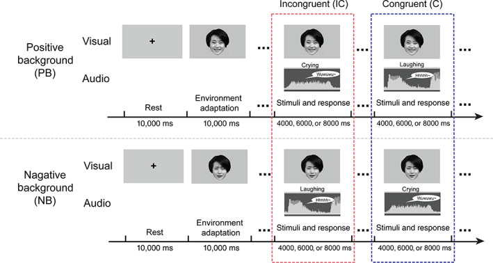
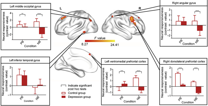
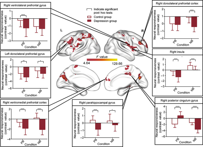
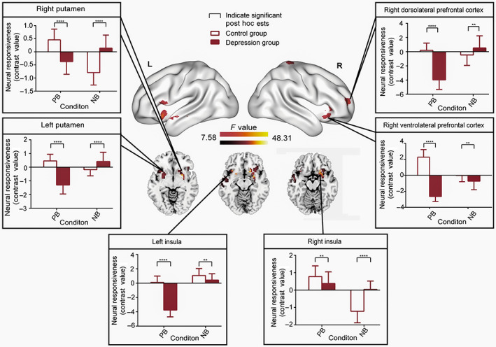
Aberrant affective neural processing and negative emotional bias are trait-marks of major depression disorders (MDDs). However, most research on biased emotional perception in depression has only focused on unimodal experimental stimuli, the neural basis of potentially biased emotional processing of multimodal inputs remains unclear. Here, we addressed this issue by implementing an audiovisual emotional task during functional MRI scanning sessions with 37 patients with MDD and 37 gender-, age- and education-matched healthy controls. Participants were asked to distinguish laughing and crying sounds while being exposed to faces with different emotional valences as background. We combined general linear model and psychophysiological interaction analyses to identify abnormal local functional activity and integrative processes during audiovisual emotional processing in MDD patients. At the local neural level, MDD patients showed increased bias activity in the ventromedial prefrontal cortex (vmPFC) while listening to negative auditory stimuli and concurrently processing visual facial expressions, along with decreased dorsolateral prefrontal cortex (dlPFC) activity in both the positive and negative visual facial conditions. At the network level, MDD exhibited significantly decreased connectivity in areas involved in automatic emotional processes and voluntary control systems during perception of negative stimuli, including the vmPFC, dlPFC, insula, as well as the subcortical regions of posterior cingulate cortex and striatum. These findings support a multimodal emotion dysregulation hypothesis for MDD by demonstrating that negative bias effects may be facilitated by the excessive ventral bottom-up negative emotional influences along with incapability in dorsal prefrontal top-down control system.
Keywords: audiovisual emotional processing; major depression disorder; negative bias effects.
© 2021 The Authors. Human Brain Mapping published by Wiley Periodicals LLC.
Conflict of interest statement
The authors declare that they have no conflict of interest.
Figures
Similar articles
-
Amygdala Activation and Connectivity to Emotional Processing Distinguishes Asymptomatic Patients With Bipolar Disorders and Unipolar Depression.Biol Psychiatry Cogn Neurosci Neuroimaging. 2019 Apr;4(4):361-370. doi: 10.1016/j.bpsc.2018.08.012. Epub 2018 Aug 31. Biol Psychiatry Cogn Neurosci Neuroimaging. 2019. PMID: 30343134
-
Crossmodal emotional integration in major depression.Soc Cogn Affect Neurosci. 2014 Jun;9(6):839-48. doi: 10.1093/scan/nst057. Epub 2013 Apr 10. Soc Cogn Affect Neurosci. 2014. PMID: 23576809 Free PMC article.
-
Integrating Multilevel Functional Characteristics Reveals Aberrant Neural Patterns during Audiovisual Emotional Processing in Depression.Cereb Cortex. 2021 Nov 23;32(1):1-14. doi: 10.1093/cercor/bhab185. Cereb Cortex. 2021. PMID: 34642754
-
The Multifaceted Role of the Ventromedial Prefrontal Cortex in Emotion, Decision Making, Social Cognition, and Psychopathology.Biol Psychiatry. 2018 Apr 15;83(8):638-647. doi: 10.1016/j.biopsych.2017.10.030. Epub 2017 Nov 20. Biol Psychiatry. 2018. PMID: 29275839 Free PMC article. Review.
-
Major depressive disorder associated alterations in the effective connectivity of the face processing network: a systematic review.Transl Psychiatry. 2024 Jan 25;14(1):62. doi: 10.1038/s41398-024-02734-0. Transl Psychiatry. 2024. PMID: 38272868 Free PMC article.
Cited by
-
Functional magnetic resonance imaging of depression: a bibliometrics and meta-analysis.Ann Gen Psychiatry. 2024 Oct 24;23(1):39. doi: 10.1186/s12991-024-00525-x. Ann Gen Psychiatry. 2024. PMID: 39449080 Free PMC article.
-
Does emotional valence affect cognitive performance and neurophysiological response during decision making? A preliminary study.Front Neurosci. 2024 Aug 9;18:1408526. doi: 10.3389/fnins.2024.1408526. eCollection 2024. Front Neurosci. 2024. PMID: 39184323 Free PMC article.
-
Altered spatio-temporal state patterns for functional dynamics estimation in first-episode drug-naive major depression.Brain Imaging Behav. 2022 Dec;16(6):2744-2754. doi: 10.1007/s11682-022-00739-1. Epub 2022 Nov 4. Brain Imaging Behav. 2022. PMID: 36333522 Free PMC article.
-
Exploring the capabilities of repetitive transcranial magnetic stimulation in major depressive disorder: Dynamic causal modeling of the neural network.Transl Psychiatry. 2025 Jul 25;15(1):257. doi: 10.1038/s41398-025-03480-7. Transl Psychiatry. 2025. PMID: 40715052 Free PMC article.
-
Declarative Memory Impairment and Emotional Bias in Recurrent Depression with a Seasonal Pattern: The Interplay between Emotion and Cognition in Seasonal Affective Disorder.Brain Sci. 2022 Oct 5;12(10):1352. doi: 10.3390/brainsci12101352. Brain Sci. 2022. PMID: 36291286 Free PMC article.
References
-
- Almeida, J. R. C. , Akkal, D. , Hassel, S. , Travis, M. J. , Banihashemi, L. , Kerr, N. , … Phillips, M. L. (2009). Reduced gray matter volume in ventral prefrontal cortex but not amygdala in bipolar disorder: Significant effects of gender and trait anxiety. Psychiatry Research ‐ Neuroimaging, 171(1), 54–68. 10.1016/j.pscychresns.2008.02.001 - DOI - PMC - PubMed
-
- Banich, M. T. , Mackiewicz, K. L. , Depue, B. E. , Whitmer, A. J. , Miller, G. A. , & Heller, W. (2009). Cognitive control mechanisms, emotion and memory: A neural perspective with implications for psychopathology. Neuroscience & Biobehavioral Reviews, 33(5), 613–630. 10.1016/j.neubiorev.2008.09.010 - DOI - PMC - PubMed
-
- Beatteay, A. , & Wilbiks, J. M. P. (2020). The effects of major depressive disorder symptoms on audiovisual integration. Journal of Cognitive Psychology, 32(8), 805–815. 10.1080/20445911.2020.1825452 - DOI
-
- Beck, A. T. (1967). Depression: Clinical, experimental and theoretical aspects. New York: Hoeber.
-
- Beck, A. T. , & Bredemeier, K. (2016). A unified model of depression. Clinical Psychological Science, 4(4), 596–619. 10.1177/2167702616628523 - DOI
Publication types
MeSH terms
LinkOut - more resources
Full Text Sources
