. 2021 Dec 10;6(66):eabj5129.
doi: 10.1126/sciimmunol.abj5129. Epub 2021 Dec 10.

Intranasal priming induces local lung-resident B cell populations that secrete protective mucosal antiviral IgA

Affiliations

Intranasal priming induces local lung-resident B cell populations that secrete protective mucosal antiviral IgA

Ji Eun Oh et al. Sci Immunol. .

Abstract

Antibodies secreted at the mucosal surface play an integral role in immune defense by serving to neutralize the pathogen and promote its elimination at the site of entry. Secretory immunoglobulin A (IgA) is a predominant Ig isotype at mucosal surfaces whose epithelial cells express polymeric Ig receptor capable of transporting dimeric IgA to the lumen. Although the role of IgA in intestinal mucosa has been extensively studied, the cell types responsible for secreting the IgA that protects the host against pathogens in the lower respiratory tract are less clear. Here, using a mouse model of influenza virus infection, we demonstrate that intranasal, but not systemic, immunization induces local IgA secretion in the bronchoalveolar space. Using single-cell RNA sequencing, we found a heterogeneous population of IgA-expressing cells within the respiratory mucosa, including tissue-resident memory B cells, plasmablasts, and plasma cells. IgA-secreting cell establishment within the lung required CXCR3. An intranasally administered protein-based vaccine also led to the establishment of IgA-secreting cells in the lung, but not when given intramuscularly or intraperitoneally. Last, local IgA secretion correlated with superior protection against secondary challenge with homologous and heterologous virus infection than circulating antibodies alone. These results provide key insights into establishment of protective immunity in the lung based on tissue-resident IgA-secreting B cells and inform vaccine strategies designed to elicit highly effective immune protection against respiratory virus infections.


Conflict of interest statement

Competing Interests A.I. is a cofounder of RIGImmune and Xanadu Bio, and serves as a consultant to 4BIO Capital, BlueWillow Biologics, Healthspan Technologies and Revelar Biotherapeutics. F.K. is listed as inventor on patent applications relating to NA-based influenza virus vaccines filed by the Icahn School of Medicine at Mount Sinai. The other authors declare that they have no competing interests.

Figures

Fig. 1.
Fig. 1.. Local but not systemic immunization with influenza A virus induces high level of IgA in local mucosa
(A) C57BL/6 mice were immunized with 20−30 pfu of influenza A/PR8 virus intranasally (i.n.) once or 4×10⁶ pfu of A/PR8 virus intraperitoneally (i.p) for 5 consecutive days. (B) Five weeks later, levels of IgG2b, IgG2c, IgA were measured from serum and bronchoalveolar lavage (BAL) fluid by ELISA (naïve, n=3; i.p. PR8, n=5; i.n. PR8, n=6). (C) Secretory IgA level in BAL fluid was measured by ELISA (naïve, n=4; i.p. PR8, n=5; i.n. PR8, n=6). Data are mean ± s.e.m. Data are representative of two independent experiments. *, p<0.05; **, p< 0.01; ***, p< 0.001
Fig. 2.
Fig. 2.. Local immunization induces IgA-producing tissue-resident cells
C57BL/6 mice were immunized with 20−30 pfu of influenza A/PR8 virus intranasally (i.n.) once or 4×10⁶ pfu of A/PR8 virus intraperitoneally (i.p) for 5 consecutive days. (A-B) Five weeks later, frozen sections of lung were stained with antibodies against B220, CD138, polymeric Ig receptor (pIgR) (red), IgA (green) and DAPI (blue). Scale bars, 100 um. (C) Five weeks later, IgA+ tissue-resident memory B cell (BRM) and antibody-secreting cells in lung were analyzed in naïve (n=2), i.p. immunized (n=4), and i.n. immunized (n=4) mice by flow cytometry. Mice were intravenously injected with anti-CD45 antibody (2 μg/mouse) 5 min prior to euthanasia to label circulating immune cells inside the blood vessels. Data are mean ± s.e.m. Data are representative of two independent experiments. *, p<0.05; **, p< 0.01; ***, p< 0.001
Fig. 3.
Fig. 3.. Mucosal IgA antibodies are secreted only after local immunization
C57BL/6 mice were immunized with 20 pfu of influenza A/PR8 virus intranasally. Eight to nine weeks later, immunized mice were surgically joined with naive mice. (A) Strategy for parabiosis. (B-C) Four to five weeks after parabiosis, flu-specific antibodies in serum (B) and BAL fluid (C) were measured by ELISA (N-N Naïve (naïve partner of naïve-naïve parabiosis), n=6; N-I Naïve (naïve partner of naïve-immunized parabiosis), n=7; N-I Immune (immunized partner of naïve-immunized parabiosis), n=7). Data are mean ± s.e.m. Data are representative of two independent experiments. *, p<0.05; **, p< 0.01; ***, p< 0.001
Fig. 4.
Fig. 4.. Single-cell sequencing reveals tissue resident IgA positive B cells localized in the lungs of immunized mice
(A) UMAP projection of tissue-resident (unresponsive to IV labeling) IgDCD19+ B cells from mediastinal lymph nodes of immunized and naïve mice. (B) UMAP projection of tissue-resident (unresponsive to IV labeling) IgDCD19+ B cells from lungs of immunized and naïve mice. (C) Gene expression heatmap of Igha and Cxcr3 projected onto UMAP. (D) Distribution of B cell clones found in plasma cells (left) and plasmablasts (right) in the lung. (E) Percent of clones found in each cluster in the lymph node (left) and lung (right) that are also found in lung plasma cells and plasmablasts. (F) GSEA plots displaying correlation of lung resident plasma cells (left), IgA+CXCR3+ B cells (middle), and plasmablasts (right) compared with their counterparts from lymph nodes to tissue resident T cells. (G) Heatmap displaying significant GSEA correlation between these cell types and listed T cell subtypes.
Fig. 5.
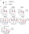
Fig. 5.. CXCR3 is required for localization of IgA+ BRM and plasma cells in the lung and IgA secretion in lung mucosa
CXCR3-B cell KO and WT control mixed bone-marrow chimeric mice were generated. Eight weeks after, mice were immunized with 25 pfu of influenza A/PR8 virus intranasally. (A) Strategy for the experiments. (B-C) Five weeks later, IgA+ plasmablasts and IgA+ plasma cells in WT (n=6) and CXCR3-B KO (n=5) mice (B) and IgA+ BRM in WT (naïve, n=4; immune, n=5), and CXCR3-B KO (naïve, n=3; immune, n=3) mice (C) were analyzed by flow cytometry. Mice were intravenously injected with anti-CD45 antibody (2 μg/mouse) 5 min prior to euthanasia to label circulating immune cells inside the blood vessels. (D) Five weeks later, levels of IgG2b, IgG2c, IgA were measured from serum and BAL fluid by ELISA (WT naïve, n=4; CXCR3-B KO naïve, n=3; WT immune, n=5; CXCR3-B KO immune, n=3). Data are mean ± s.e.m. Data are representative of two independent experiments. *, p<0.05; ***, p< 0.001
Fig. 6.
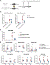
Fig. 6.. Local vaccination with adjuvanted recombinant neuraminidase induces IgA secretion in lung
(A) C57BL/6 mice were vaccinated with 10 μg recombinant neuraminidase (rNA) from A/Michigan/15/45 adjuvanted with 5 μg poly (I:C) intranasally (i.n.), intramuscularly (i.m.), or intraperitoneally (i.p), and boosted with same agents 3–4 weeks later. (B) Four weeks later, levels of NA-specific IgG2b, IgG2c, IgA were measured from serum and BAL fluid by ELISA (n=4). Data are mean ± s.e.m. Data are representative of two independent experiments. *, p<0.05; **, p< 0.01
Fig. 7.
Fig. 7.. Locally immunized mice show superior protection against secondary challenge
(A-D) C57BL/6 mice were immunized with 20 pfu of influenza A/PR8 virus intranasally once or 4×10⁶ pfu intraperitoneally for 5 consecutive days. Five weeks later, CD4 T and CD8 T cells were depleted intravenously with 300 μg each of anti-CD4 (GK1.5) and anti-CD8 (2.43) antibodies on d−4, d−1, d+2, and d+4 post challenge and intranasally with 100 μg each of anti-CD4 and anti-CD8 antibodies on d−1 post challenge. (A-B) Mice were challenged intranasally with a high dose of A/PR8 virus (2×10⁶ pfu). (A) Body weights were monitored (naïve, n=6; immunized, n=5). (B) Virus titer in BAL fluid and lung tissue lysates was analyzed at the indicated days post challenge (n=3 per group). Virus titer in two out of three lysates of i.n. PR8 group at day 2 post challenge was not detected. Dashed line indicates the limit of detection. (C-D) Mice were challenged intranasally with X31-OVA (2×10⁴ pfu) virus. (C) Body weight were monitored (naïve, n=6; immunized, n=7). (D) The levels of X31-specific IgA or IgG from BAL fluid and serum at the indicated days post challenge were measured by ELISA (n=3 per group). Sample dilution for ELISA was 1:5 (BAL IgA), 1:10 (BAL IgG) or 1:103 (Serum IgG). (E) CXCR3-B cell KO and WT control mixed bone-marrow chimeric mice were immunized with 25 pfu of influenza A/PR8 virus intranasally. Five weeks later, CD4 T and CD8 T cells were depleted intravenously with 300 μg each of anti-CD4 (GK1.5) and anti-CD8 (2.43) antibodies on d−4, d−1, d+2, and d+4 post challenge and intranasally with 100 μg each of anti-CD4 and anti-CD8 antibodies on d−1 post challenge. Mice were challenged intranasally with high dose of A/PR8 virus (2×10⁶ pfu). Body weight were monitored (naïve, n=3; immune, n=5). (F) Mice were vaccinated with 10 μg recombinant neuraminidase (rNA) from A/Michigan/15/45 adjuvanted with 5 μg poly (I:C) intranasally (i.n.), intramuscularly (i.m.), or intraperitoneally (i.p), and boosted with same agents 3 weeks later. CD4 T and CD8 T cells were depleted intravenously with 300 μg each of anti-CD4 (GK1.5) and anti-CD8 (2.43) antibodies on d−4, d−1, d+2, and d+4 post challenge and intranasally with 100 μg each of anti-CD4 and anti-CD8 antibodies on d-1 post challenge. Mice were challenged intranasally with lethal dose of A/PR8 virus (100 pfu). Body weight and survival were monitored (naïve, n=5; i.n., n=7; i.m., n=6; i.p., n=5). Data are mean ± s.e.m. Data are representative of two independent experiments or are pooled from two independent experiments (C). *, p<0.05; **, p< 0.01; ***, p< 0.001; ****, p< 0.0001

References

    1. Iwasaki A, Exploiting Mucosal Immunity for Antiviral Vaccines. Annu Rev Immunol 34, 575–608 (2016). - PubMed
    1. Allie SR, Bradley JE, Mudunuru U, Schultz MD, Graf BA, Lund FE, Randall TD, The establishment of resident memory B cells in the lung requires local antigen encounter. Nat Immunol 20, 97–108 (2019). - PMC - PubMed
    1. Corthesy B, Multi-faceted functions of secretory IgA at mucosal surfaces. Front Immunol 4, 185 (2013). - PMC - PubMed
    1. Macpherson AJ, Yilmaz B, Limenitakis JP, Ganal-Vonarburg SC, IgA Function in Relation to the Intestinal Microbiota. Annu Rev Immunol 36, 359–381 (2018). - PubMed
    1. Kaetzel CS, Robinson JK, Lamm ME, Epithelial transcytosis of monomeric IgA and IgG cross-linked through antigen to polymeric IgA. A role for monomeric antibodies in the mucosal immune system. J Immunol 152, 72–76 (1994). - PubMed

Publication types

MeSH terms