. 2022;13(3):925-947.
doi: 10.1016/j.jcmgh.2021.12.002. Epub 2021 Dec 8.

Sphingosine 1-Phosphate Receptor 4 Promotes Nonalcoholic Steatohepatitis by Activating NLRP3 Inflammasome

Affiliations

Sphingosine 1-Phosphate Receptor 4 Promotes Nonalcoholic Steatohepatitis by Activating NLRP3 Inflammasome

Chung Hwan Hong et al. Cell Mol Gastroenterol Hepatol. 2022.

Abstract

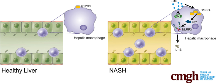
Background & aims: Sphingosine 1-phosphate receptors (S1PRs) are a group of G-protein-coupled receptors that confer a broad range of functional effects in chronic inflammatory and metabolic diseases. S1PRs also may mediate the development of nonalcoholic steatohepatitis (NASH), but the specific subtypes involved and the mechanism of action are unclear.

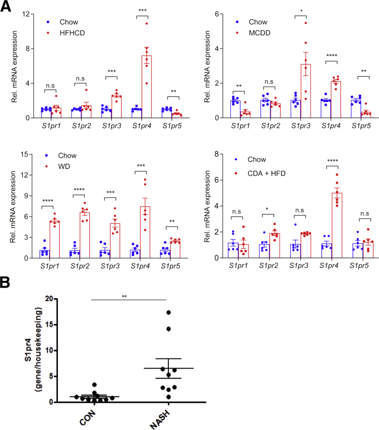
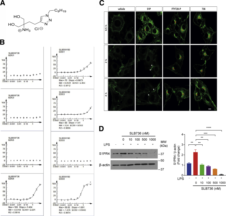
Methods: We investigated which type of S1PR isoforms is activated in various murine models of NASH. The mechanism of action of S1PR4 was examined in hepatic macrophages isolated from high-fat, high-cholesterol diet (HFHCD)-fed mice. We developed a selective S1PR4 functional antagonist by screening the fingolimod (2-amino-2-[2-(4- n -octylphenyl)ethyl]-1,3- propanediol hydrochloride)-like sphingolipid-focused library.

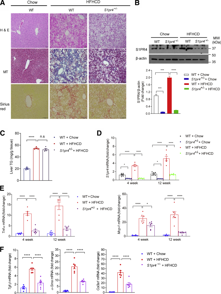
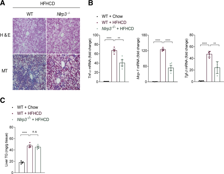
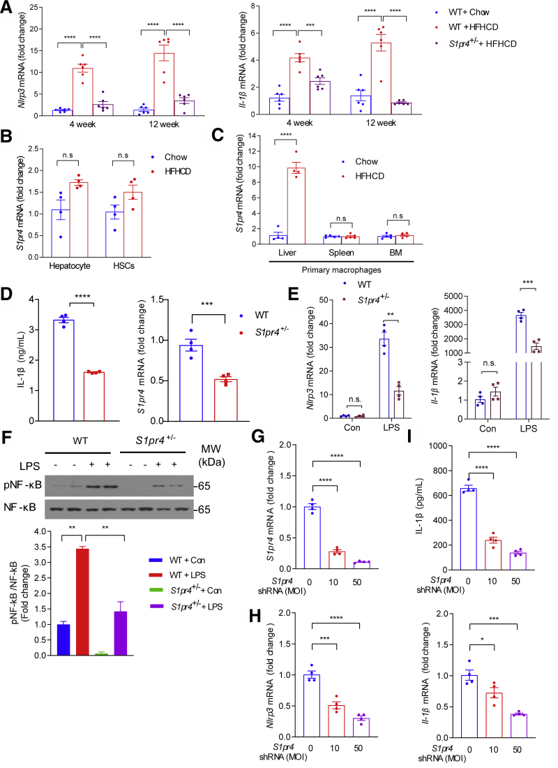
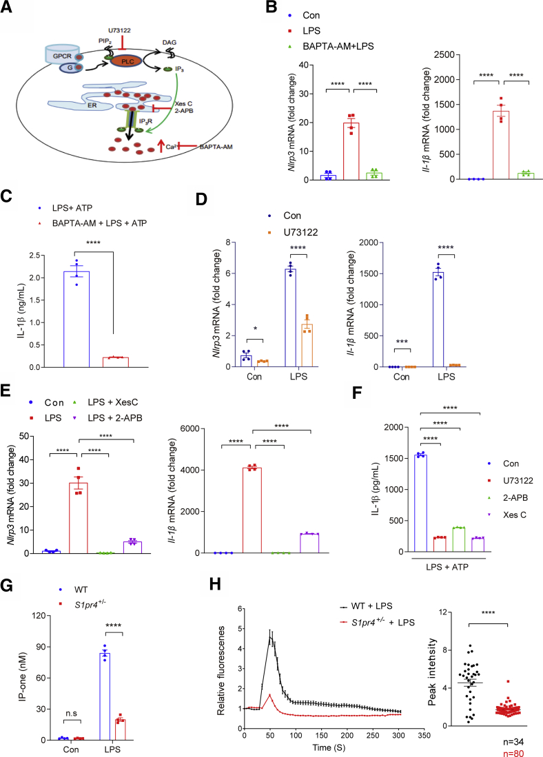
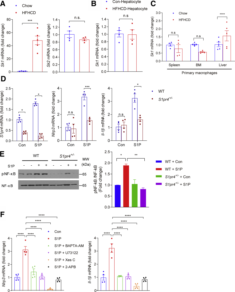
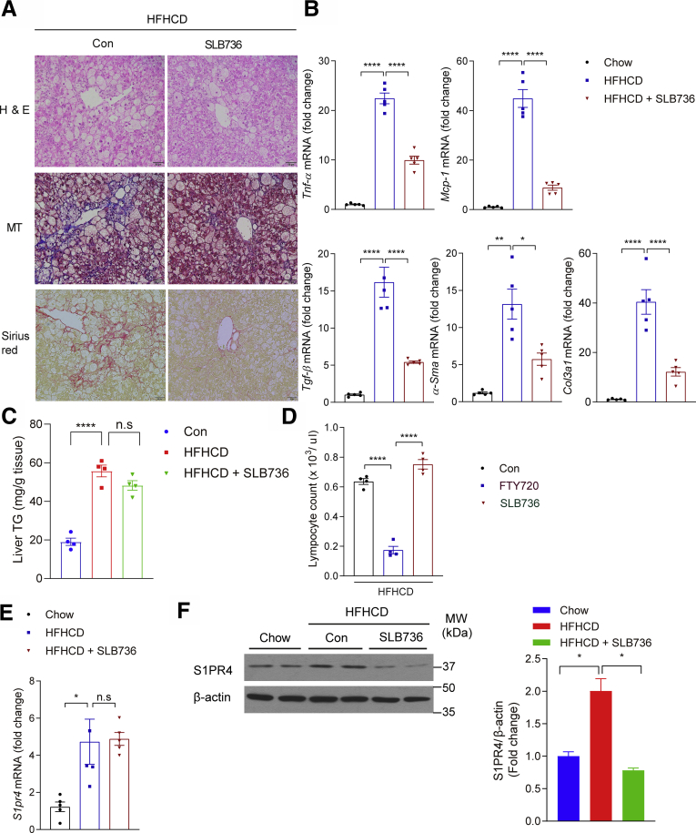
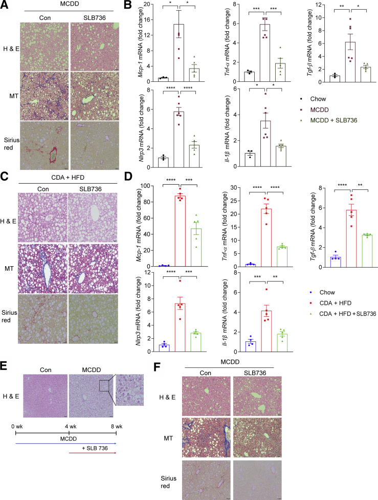
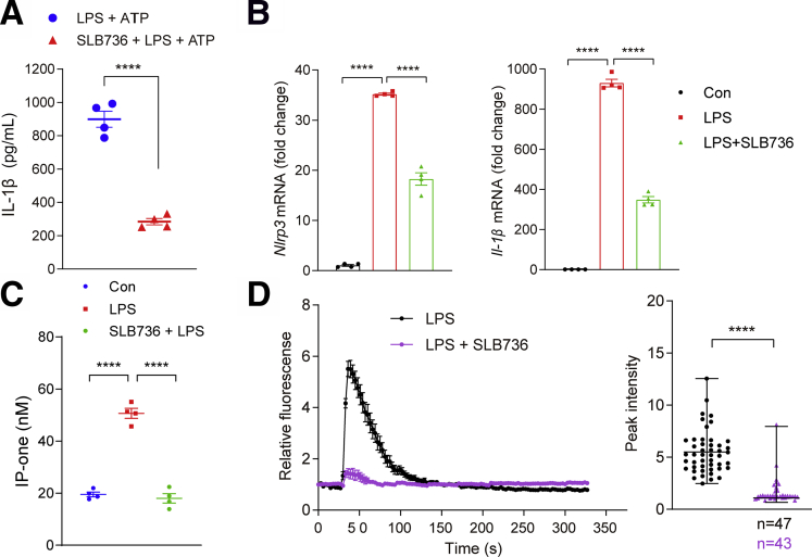
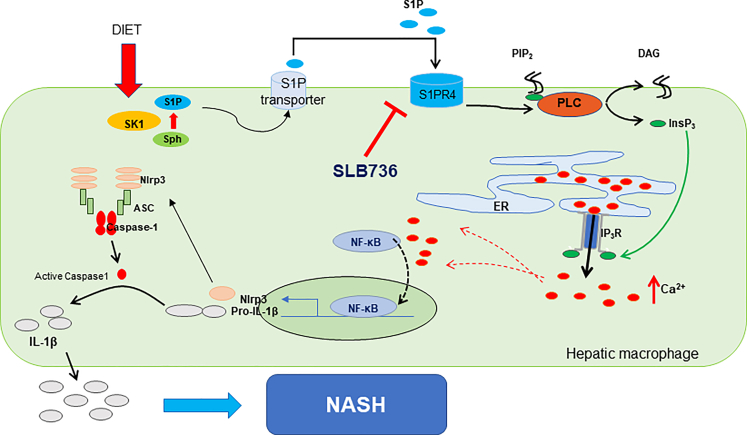
Results: The livers of various mouse models of NASH as well as hepatic macrophages showed high expression of S1pr4. Moreover, in a cohort of NASH patients, expression of S1PR4 was 6-fold higher than those of healthy controls. S1pr4+/- mice were protected from HFHCD-induced NASH and hepatic fibrosis without changes in steatosis. S1pr4 depletion in hepatic macrophages inhibited lipopolysaccharide-mediated Ca++ release and deactivated the Nod-like receptor pyrin domain-containning protein 3 (NLRP3) inflammasome. S1P increased the expression of S1pr4 in hepatic macrophages and activated NLRP3 inflammasome through inositol trisphosphate/inositol trisphosphate-receptor-dependent [Ca++] signaling. To further clarify the biological function of S1PR4, we developed SLB736, a novel selective functional antagonist of SIPR4. Similar to S1pr4+/- mice, administration of SLB736 to HFHCD-fed mice prevented the development of NASH and hepatic fibrosis, but not steatosis, by deactivating the NLRP3 inflammasome.

Conclusions: S1PR4 may be a new therapeutic target for NASH that mediates the activation of NLRP3 inflammasome in hepatic macrophages.

Keywords: Ca(++); Functional Antagonist; Hepatic Macrophages; S1P.


Figures

None
Graphical abstract
Figure 1
Figure 1
Hepatic S1pr4 expression is uniquely increased in the liver of murine NASH models. (A) Diet was administered to mice for 12 weeks for HFHCD, 8 weeks for MCDD, 16 weeks for WD, and 6 weeks for CDA+HFD. (A) Hepatic S1pr mRNA expression in HFHCD-, MCDD-, WD-, and CDA+HFD–induced dietary models of NASH (n = 6). (B) Hepatic mRNA expression levels of S1PR4 in the liver samples of patients with NASH/cirrhosis undergoing liver transplantation (n = 9). Surgical specimens of the donor livers were used as controls (n = 10). All data are shown as means ± SEM. Data were analyzed by Student two-tailed unpaired t test. ∗P < .05, ∗∗P < .01, ∗∗∗P < .001, and ∗∗∗∗P < .0001. CON, control; Rel., relative.
Figure 2
Figure 2
S1PR4 is a critical mediator of hepatic inflammation and fibrosis.S1pr4+/- mice were fed a chow diet or HFHCD for 12 weeks. (A) Representative H&E, Masson's trichrome, and Sirius Red staining in liver tissues. Scale bar: 50 μm. (B) Hepatic S1PR4 protein expression in the S1pr4+/- mice fed with HFHCD (n = 6). The S1PR4 protein was normalized to β-actin. (C) Liver TG content (n = 4). (D) Hepatic S1pr4 mRNA expression and (E) relative mRNA expression levels of the genes associated with inflammation (Tnf-α, Mcp-1) (n = 6). S1pr4+/- mice were fed chow or HFHCD for 4 or 12 weeks. (F) Relative mRNA expression of fibrosis (Tgf-β, α-Sma, Col3al) (n = 6). S1pr4+/- mice were fed a chow diet or HFHCD for 12 weeks. All data are shown as means ± SEM. (B–F) Data were analyzed by one-way analysis of variance with Bonferroni correction. ∗P < .05, ∗∗∗P < .0005, and ∗∗∗∗P < .0001. MW, molecular weight.
Figure 3
Figure 3
Nlrp3-/-mice are protected from the development of NASH. (A) Representative H&E and Masson's trichrome staining of the livers of WT and Nlrp3-/- mice fed HFHCD for 12 weeks. Scale bar: 50 μm. (B) Relative mRNA expression levels of the genes associated with inflammation and fibrosis in the liver (n = 4). (C) TG levels in the livers of WT mice fed chow or HFHCD, and Nlrp3-/- mice fed HFHCD (n = 4). All data are shown as means ± SEM. (B and C) Data were analyzed by one-way analysis of variance with Bonferroni correction. ∗∗P < .01, ∗∗∗∗P < .0001.
Figure 4
Figure 4
S1PR4 depletion decreases NLRP3 inflammasome activation in hepatic macrophages. (A) Relative hepatic mRNA expression levels of the genes associated with the components of NLRP3 inflammasome (n = 6). S1pr4+/- mice were fed chow or HFHCD for 4 or 12 weeks. (B) mRNA expression of S1pr4 in primary hepatocytes and HSCs from mice fed chow diet or HFHCD for 4 weeks (n = 4). (C) mRNA expression of S1pr4 in macrophages isolated from the liver, spleen, and BM. S1pr4+/- mice were fed chow or HFHCD for 4 weeks (n = 4). (D–F) S1pr4 depletion decreases NLRP3 inflammasome activation in hepatic macrophages. (D) Hepatic macrophages isolated from S1pr4+/- mice were stimulated with LPS (100 ng/mL) for 3 hours followed by ATP (1 mmol/L) for 30 minutes. Cell culture media were collected and IL-1β levels were measured by ELISA (n = 4). (E) Relative mRNA expression levels of Nlrp3 and Il-1β (n = 4) and (F) representative Western blots of NF-κB phosphorylation (pNF-κB) and corresponding quantification (n = 3). S1pr4+/- hepatic macrophages were stimulated with LPS (100 ng/mL) for (E) 3 hours or (F) 30 minutes. (G–I) Dose-dependent effect of S1pr4 shRNA transfection on the NLRP3 inflammasome in hepatic macrophages. Relative mRNA expression levels of (G) S1pr4 and (H) Nlrp3 and Il-1β are shown. (I) IL-1β in the cell culture media after transfection of normal hepatic macrophages with the indicated multiplicity of infection (MOI) of S1pr4 shRNA. Hepatic macrophages were infected with lentiviral vectors coding S1pr4 shRNA at an MOI of 0, 10, and 50. After 48 hours, hepatic macrophages were stimulated with (G and H) LPS (100 ng/mL) for 3 hours, (I) followed by stimulation with ATP (1 mmol/L) for 30 minutes. All data are shown as means ± SEM. (A and F–I) Data were analyzed by one-way analysis of variance with Bonferroni correction. (B–E) Data were analyzed by Student two-tailed unpaired t test. ∗P < .05, ∗∗P < .01, ∗∗∗P < .001, and ∗∗∗∗P < .0001. Con, control; MW, molecular weight.
Figure 5
Figure 5
Intracellular calcium signaling is necessary for S1PR4-dependent activation of NLRP3 inflammasome in hepatic macrophages. (A) Schematic illustration of inhibitor of PLC and IP3. (B and C) Effect of calcium depletion on LPS-induced activation of NLRP3 inflammasome. Relative mRNA expression levels of (B) Nlrp3 and Il- in the cells and (C) IL-1β secretion into the culture medium (n = 4). (B) Hepatic macrophages were pretreated with BAPTA-AM (10 μmol/L) for 30 minutes and then treated with LPS (100 ng/mL) for 3 hours. (C) In another set of experiments, LPS-primed hepatic macrophages were treated with ATP (1 mmol/L) for 30 minutes. (D–F) Inhibition of LPS-induced NLRP3 inflammasome pathway by suppression of the PLC/IP3/IP3R axis. (D) Relative mRNA expression levels of Nlrp3 and Il-1β. Hepatic macrophages were treated with (D) 10 μmol/L U73122, (E) 5 μmol/L Xes C or 100 μmol/L 2-APB (n = 4). (F) IL-1β in the culture medium. Hepatic macrophages pretreated with U73122 (10 μmol/L), Xes C (5 μmol/L), or 2-APB (100 μmol/L) for 30 minutes and dimethyl sulfoxide as vehicle control. Hepatic macrophages were treated with 1 mmol/L ATP for 30 minutes after 3 hours of LPS priming (100 ng/mL). IL-1β secreted in the culture supernatants was quantified by ELISA (n = 4). (G and H) S1PR4-dependent calcium release from ER plays a pivotal role in the priming of NLRP3 inflammasome in hepatic macrophages. (G) Levels of IP-one in S1pr4+/- hepatic macrophages (n = 4). (H) Effect of S1PR4 on LPS-mediated [Ca++] release. WT or S1pr4+/- hepatic macrophages were incubated with Fluo-4/AM followed by stimulation with LPS for 2 hours. [Ca++] was analyzed by time-lapse confocal microscopy (left panel). Quantification of LPS-induced peak fluorescent intensities (right panel). All data are shown as means ± SEM. (C, D, G, and H) Data were analyzed by Student two-tailed unpaired t test. (B, E, and F) Data were analyzed by one-way analysis of variance with Bonferroni correction. ∗P < .05, ∗∗∗P < .001, and ∗∗∗∗P < .0001. Con, control; DAG, diacylglycerol; G, G-protein; GPCR, G protein-coupled receptor.
Figure 6
Figure 6
S1P activates NLRP3 inflammasome through S1PR4. (A) Sk1 and Sk2 mRNA expression in the livers of mice fed HFHCD for 12 weeks (n = 4). (B and C) Sk1 mRNA expression in (B) primary hepatocytes and (C) in macrophages from mice fed HFHCD for 4 weeks (n = 4–5). Macrophages were isolated from the liver, spleen, and BM. (D and E) S1pr4 knockdown decreases the S1P-induced NLRP3 inflammasome activation. (D) The mRNA expression of S1pr4, Nlrp3, and Il-. Serum-starved S1pr4+/- hepatic macrophages were treated with S1P (1 μmol/L) for 2 hours (n = 4). (E) Representative Western blots of NF-κB phosphorylation (pNF-κB) and corresponding quantification (n = 3). S1pr4+/- hepatic macrophages were treated with S1P (1 μmol/L) for 30 minutes. (F) mRNA expression of Nlrp3 and Il-. Serum-starved hepatic macrophages were treated with BAPTA-AM (10 μmol/L), U73122 (10 μmol/L), Xes C (5 μmol/L), or 2-APB (100 μmol/L), and treated with S1P (1 μmol/L) (n = 4). All data are shown as means ± SEM. (AD) Data were analyzed by Student two-tailed unpaired t test. (E and F) Data were analyzed by one-way analysis of variance followed by Bonferroni correction. ∗P < .05, ∗∗P < .01, ∗∗∗P < .001, and ∗∗∗∗P < .0001. Con, control; MW, molecular weight.
Figure 7
Figure 7
SLB736 acts as a functional antagonist of S1PR4. (A) Structure of SLB736. (B) Isoform-specific S1PR activity of SLB736. β-arrestin PathHunter assay was performed in the clonal S1PR/HEK293 cell line in the presence of SLB736 (left) and FTY720-P (right). EDG 1 = S1PR1, EDG5 = S1PR2, EDG3 = S1PR3, EDG6 = S1PR4. (C) S1PR4 internalization and recycling were assessed in C6 glioma cells overexpressing EGFP-fused S1PR4. Cells were exposed to vehicle (0.1% BSA), S1P (100 nmol/L), FTY720-P (1 μmol/L), or SLB736 (1 μmol/L) for 0.5 hours and fixed. Cells exposed to reagents for 0.5 hours were washed and further incubated with vehicle in the presence of cyclohexamide for up to 4 hours. Asterisks or arrowheads indicate cytosolic locations or the cell surface. Scale bar: 10 μm. (D) Protein level of S1PR4 in hepatic macrophages. The S1PR4 protein was normalized to β-actin. Hepatic macrophages were pretreated with SLB736 at the indicated dose and then treated with 100 ng/mL of LPS for 24 hours (n = 4). All data are shown as means ± SEM. (D) Data were analyzed by one-way analysis of variance followed by Bonferroni correction. ∗∗P < .01, ∗∗∗P < .001, and ∗∗∗∗P < .0001. EC50, 50% Effective Concentration; Max, maximum; Min, minimum; MW, molecular weight.
Figure 8
Figure 8
SLB736 treatment prevents HFHCD-induced NASH. (A) Representative H&E, Masson's trichrome, and Sirius Red staining of the livers. Scale bars: 50 μm. SLB736 (1 mg/kg/d) was administered for 12 weeks in HFHCD-fed mice. (B) Relative mRNA expression levels of the genes associated with inflammation (Tnf-α, Mcp-1) and fibrosis (Tgf-β, α-Sma, Col3al) (n = 5). (C) Liver TG contents (n = 4). (D) Lymphocyte counts in the blood. FTY720 or SLB736 (1 mg/kg/d) were administered for 12 weeks in HFHCD-fed mice (n = 4). (E and F) mRNA expression of (E) S1pr4 and (F) representative Western blots of S1PR4 and corresponding quantification in the liver of HFHCD-fed mice treated with SLB736 (n = 4). All data are shown as means ± SEM. (BF) Data were analyzed by one-way analysis of variance followed by Bonferroni correction. ∗P < .05, ∗∗P < .01, and ∗∗∗∗P < .0001. Con, control; MW, molecular weight.
Figure 9
Figure 9
SLB736 prevents NASH in other animal models and slowsthe progression to NASH and fibrosis. (A) Representative H&E, Masson's trichrome, and Sirius Red staining of the livers of MCDD-fed mice with or without SLB736 treatment for 8 weeks. Scale bars: 50 μm. (B) Relative mRNA expression levels of inflammation, fibrosis, and inflammasome markers in the livers of chow-fed mice and MCDD-fed mice with or without SLB736 (n = 3–6). (C) Representative H&E, MT, and Sirius Red staining of the livers of CDA+HFD–fed mice with or without SLB736 treatment for 6 weeks. Scale bars: 50 μm. (D) Relative mRNA expression levels of markers for inflammation, fibrosis, and inflammasome in the livers of chow-fed mice and CDA+HFD–fed mice with or without SLB736 treatment (n = 4–5). (E) Representative H&E of livers of MCDD-fed mice for 4 weeks. Scale bars: 50 μm. Inlet shows the lipid accumulation in hepatocytes. Lower panel: Schematic schedule for observing the therapeutic effect of SLB736 in MCDD-fed mice. (F) Representative H&E, MT, and Sirius Red staining of the livers. Scale bars: 50 μm. After feeding MCDD for 4 weeks, SLB736 (1 mg/kg/d) was administrated for 4 weeks with MCDD. All data are shown as means ± SEM. (B and D) Data were analyzed by one-way analysis of variance followed by Bonferroni correction. ∗P < .05, ∗∗P < .01, ∗∗∗P < .001, and ∗∗∗∗P < .0001. Con, control.
Figure 10
Figure 10
SLB736 decreases NLRP3 inflammasome activation in hepatic macrophages. (A) IL-1β levels in the culture media of hepatic macrophages. Hepatic macrophages were pretreated with 1 μmol/L SLB736 for 2 hours, and treated with LPS 100 ng/mL for 3 hours followed by ATP (1 mmol/L) for 30 minutes. (B) Relative mRNA expression of Nlrp3 and Il-. Hepatic macrophages were pretreated with 1 μmol/L SLB736 for 2 hours, and treated with LPS 100 ng/mL for 3 hours (n = 4). (C) IP-one levels in hepatic macrophages. Cells were pretreated with 1 μmol/L SLB736 or vehicle for 2 hours (n = 4). The levels of IP-one in cell lysates was measured by an ELISA kit. (D) Effect of SLB736 on LPS-mediated [Ca++] release. Hepatic macrophages were pretreated with 1 μmol/L SLB736 or vehicle for 2 hours. Cells then were incubated with Fluo-4/AM followed by stimulation with LPS. [Ca++] was analyzed by time-lapse confocal microscopy (left panel). Quantification of LPS-induced peak fluorescent intensities (right panel). All data are shown as means ± SEM. (A and D) Data were analyzed by Student two-tailed unpaired t test. (B and C) Data were analyzed by one-way analysis of variance with Bonferroni correction. ∗∗∗∗P < .0001. Con, control.
Figure 11
Figure 11
Conceptual model showing the role of the SK1/S1PR4 axis in the pathogenesis of NASH. S1P produced by SK1 from hepatic macrophages induces S1PR4 in a paracrine manner, which is necessary for the [Ca++]-dependent priming of the NLRP3 inflammasome. ASC, Apoptosis-associated speck-like protein containing a C-terminal caspase-recruitment domain; DAG, diacylglycerol; Sph, sphingosine.

References

    1. Friedman S.L., Neuschwander-Tetri B.A., Rinella M., Sanyal A.J. Mechanisms of NAFLD development and therapeutic strategies. Nat Med. 2018;24:908–922. - PMC - PubMed
    1. Lassailly G., Caiazzo R., Pattou F., Mathurin P. Perspectives on treatment for nonalcoholic steatohepatitis. Gastroenterology. 2016;150:1835–1848. - PubMed
    1. Younossi Z.M., Loomba R., Rinella M.E., Bugianesi E., Marchesini G., Neuschwander-Tetri B.A., Serfaty L., Negro F., Caldwell S.H., Ratziu V., Corey K.E., Friedman S.L., Abdelmalek M.F., Harrison S.A., Sanyal A.J., Lavine J.E., Mathurin P., Charlton M.R., Chalasani N.P., Anstee Q.M., Kowdley K.V., George J., Goodman Z.D., Lindor K. Current and future therapeutic regimens for nonalcoholic fatty liver disease and nonalcoholic steatohepatitis. Hepatology. 2018;68:361–371. - PMC - PubMed
    1. Machado M.V., Diehl A.M. Pathogenesis of nonalcoholic steatohepatitis. Gastroenterology. 2016;150:1769–1777. - PMC - PubMed
    1. Koh E.H., Yoon J.E., Ko M.S., Leem J., Yun J.Y., Hong C.H., Cho Y.K., Lee S.E., Jang J.E., Baek J.Y., Yoo H.J., Kim S.J., Sung C.O., Lim J.S., Jeong W.I., Back S.H., Baek I.J., Torres S., Solsona-Vilarrasa E., Conde de la Rosa L., Garcia-Ruiz C., Feldstein A.E., Fernandez-Checa J.C., Lee K.U. Sphingomyelin synthase 1 mediates hepatocyte pyroptosis to trigger non-alcoholic steatohepatitis. Gut. 2021;70:1954–1964. - PMC - PubMed

Publication types

Substances