. 2022 Jan:280:121270.
doi: 10.1016/j.biomaterials.2021.121270. Epub 2021 Nov 22.

A poly(ethylene glycol) three-dimensional bone marrow hydrogel

Affiliations

A poly(ethylene glycol) three-dimensional bone marrow hydrogel

Lauren E Jansen et al. Biomaterials. 2022 Jan.

Abstract

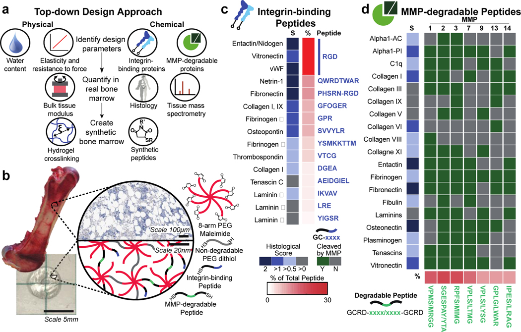
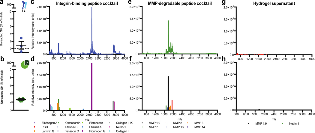
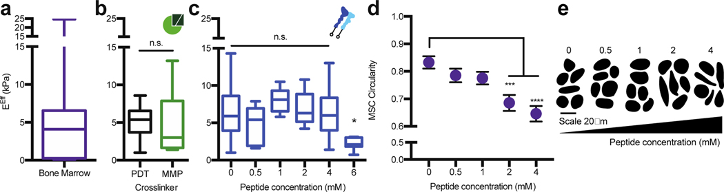
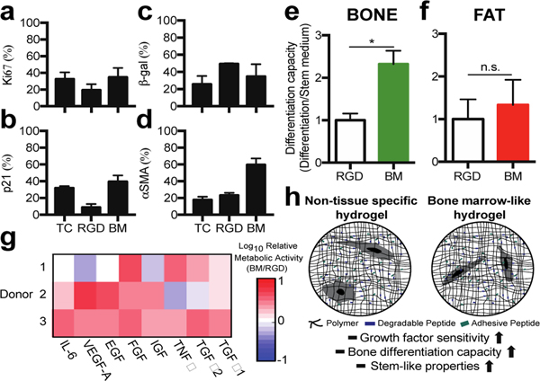
Three-dimensional (3D) hydrogels made from synthetic polymers have emerged as in vitro cell culture platforms capable of representing the extracellular geometry, modulus, and water content of tissues in a tunable fashion. Hydrogels made from these otherwise non-bioactive polymers can be decorated with short peptides derived from proteins naturally found in tissues to support cell viability and direct phenotype. We identified two key limitations that limit the ability of this class of materials to recapitulate real tissue. First, these environments typically display between 1 and 3 bioactive peptides, which vastly underrepresents the diversity of proteins found in the extracellular matrix (ECM) of real tissues. Second, peptides chosen are ubiquitous in ECM and not derived from proteins found in specific tissues, per se. To overcome this critical limitation in hydrogel design and functionality, we developed an approach to incorporate the complex and specific protein signature of bone marrow into a poly (ethylene glycol) (PEG) hydrogel. This bone marrow hydrogel mimics the elasticity of marrow and has 20 bone marrow-specific and cell-instructive peptides. We propose this tissue-centric approach as the next generation of 3D hydrogel design for applications in tissue engineering and beyond.

Keywords: 3D biomaterial; Mesenchymal stem cell; Peptide; Stiffness; Tissue mimic; integrin.


Figures

Figure 1.
Figure 1.. A PEG hydrogel designed to mimic the physical and chemical properties of bone marrow tissue.
a) Tissues have specific physical and chemical properties such as water content, elasticity, integrin-binding, and MMP-degradable proteins. These properties can be quantified in real bone marrow tissue using rheology, mass spectrometry, and tissue histology (Image of human adapted from Protein Atlas). In PEG hydrogels, these features can be mimicked by tuning the polymer crosslinking density and incorporating peptides (histology from the Protein Atlas). b) Here, bone marrow tissue (image of porcine bone marrow) is mimicked with a hydrogel composed of an 8-arm PEG macromer functionalized (image of resulting hydrogel) with c) 13 cysteine-terminated integrin-binding peptides, and crosslinked with d) 7 di-cysteine-terminated MMP-degradable peptides and PEG-dithiol. The known functional sequence for each peptide is depicted in blue for integrin-binding proteins (up to the first 8 amino acids are depicted) and in green for the degradable peptides, where the slash (/) indicates the cleavage location for each enzyme on the matched peptide. Scales for the average histological score and the total percent of each peptide are shown by each peptide/protein pair (Y=yes, N=no, S=Histological Score).
Figure 2.
Figure 2.. Validation of bone marrow hydrogel peptides.
a) Cells were treated with peptides in solution (medium), and then seeded onto coverslips coated with the bone marrow integrin-binding peptide cocktail. MSC area was measured over approximately 2 hours for cells not treated (control, black) or pre-treated for 30 minutes prior (blue) with soluble integrin-binding peptides and allowed to adhere to a coverslip coupled with all the integrin-binding peptides included in the bone marrow hydrogel design. Representative cell images (scale bar = 50 μm) and traces of MSCs 2 hours after seeding (bottom). Error bars represent SEM. b) Heat map depicting the log10 fold change in cell area at 2 hours compared to no treatment (NT) for each integrinbinding peptide for hTERT MSCs (hT) and three donor MSCs (1–3) (BM=bone marrow peptide cocktail) (N≥2, n≥20 per cell). c) Representative image of MSCs seeded on cytodex beads (black outline) and encapsulated into a hydrogel with MMP degradable crosslinkers (Cell area=red, branch length=green). d) A box and whisker plot for the maximum branch length per bead in each hydrogel condition. e) Representative cell and bead traces in each hydrogel condition, where the lighter colored circle is the bead and the darker color is the cell trace (N=2, n≥15 per cell). Significance is determined using a two-tailed t-test. P-values <0.05 are considered significant, where p<0.05 is denoted with *, ≤0.01 with **, ≤0.001 with ***, and ≤0.0001 with ****.
Figure 3.
Figure 3.. Bone marrow peptides couple to the hydrogel at expected concentrations.
a) The percentage of unreacted thiols when integrin-binding peptides were added to a solution of PEG-maleimide dissolved in PBS at pH 7.4. b) The percentage of unreacted thiols 10 minutes post-crosslinking an 8-arm PEG hydrogel at a 1:1 molar ratio of thiol to maleimide. Error bars represent the SEM (N≥1, n≥3). MALDI-TOF spectrum (top) and identified peptide peaks (bottom) for the c) and d) bone marrow integrin-binding peptides; e) and f) the bone marrow MMP-sensitive peptide crosslinkers, and g) and h) the supernatant of a bone marrow hydrogel swelled for 4 hours in PBS.
Figure 4.
Figure 4.. The PEG hydrogel accurately models the bulk compressive properties of bone marrow tissue.
a) Rheology data from Jansen et al., 20151 for the effective Young’s modulus (EEff) of porcine bone marrow at 35°C. b) The EEff for 20 wt%, 8-arm, 20K PEG hydrogels crosslinked at a 1:1 thiol to maleimide molar ratio with 1.5 kDa PEG-dithiol (PDT, black) or with the bone marrow cocktail containing MMP crosslinkers (MMP, green). c) The EEff for 20 wt%, 8-arm, 20K PEG hydrogels crosslinked at a 1:1 thiol to maleimide molar ratio with PDT and coupled with different concentrations of the bone marrow peptide cocktail for 10 minutes before gelation. d) MSCs circularity with respect to peptide concentration and e) representative cell traces for cells encapsulated in a 20 wt%, 8-arm, 20 kDa PEG-crosslinked with the bone marrow cocktail. The significance is determined using a two-tailed t-test where p=0.05, and error bars represent the SEM. (N≥2, n≥3 for mechanical testing; N≥2, n≥10 for cell circularity).
Figure 5.
Figure 5.. The bone marrow hydrogel supports MSC growth, and stem-like properties.
Staining for a) Ki67, b) p21, c) beta-galactosidase, and d) α-smooth muscle actin positive cells in a hydrogel with no degradability and 2 mM RGD (RGD) or the bone marrow hydrogel (BM). e) Oil Red O or f) Osteoimage differentiation capacity normalized to the RGD hydrogel. g) Log10 of cell metabolic activity three days after cell encapsulation into the bone marrow hydrogel or an RGD hydrogel for all donor MSCs. Each growth factor was dosed at 20 ng/mL in cell culture medium (n≥3). h) Schematic to compare how the two hydrogels impact observed MSC phenotypes.

References

    1. Jansen LE, Birch NP, Schiffman JD, Crosby AJ & Peyton SR Mechanics of intact bone marrow. J Mech Behav Biomed Mater 50, 299–307, doi:10.1016/j.jmbbm.2015.06.023 (2015). - DOI - PMC - PubMed
    1. Nguyen TV, Sleiman M, Moriarty T, Herrick WG & Peyton SR Sorafenib resistance and JNK signaling in carcinoma during extracellular matrix stiffening. Biomaterials 35, 5749–5759, doi:10.1016/j.biomaterials.2014.03.058 (2014). - DOI - PubMed
    1. Barney LE et al. A cell-ECM screening method to predict breast cancer metastasis. Integr Biol (Camb) 7, 198–212, doi:10.1039/c4ib00218k (2015). - DOI - PMC - PubMed
    1. Herrick WG et al. PEG-phosphorylcholine hydrogels as tunable and versatile platforms for mechanobiology. Biomacromolecules 14, 2294–2304, doi:10.1021/bm400418g (2013). - DOI - PubMed
    1. Uhlén M. et al. Tissue-based map of the human proteome. Science 347, 1260419 (2015). - PubMed

Publication types

MeSH terms