Review
. 2022 Jan 17;23(1):bbab504.
doi: 10.1093/bib/bbab504.

Tumor immune microenvironment lncRNAs

Affiliations
Review

Tumor immune microenvironment lncRNAs

Eun-Gyeong Park et al. Brief Bioinform. .

Abstract

Long non-coding ribonucleic acids (RNAs) (lncRNAs) are key players in tumorigenesis and immune responses. The nature of their cell type-specific gene expression and other functional evidence support the idea that lncRNAs have distinct cellular functions in the tumor immune microenvironment (TIME). To date, the majority of lncRNA studies have heavily relied on bulk RNA-sequencing data in which various cell types contribute to an averaged signal, limiting the discovery of cell type-specific lncRNA functions. Single-cell RNA-sequencing (scRNA-seq) is a potential solution for tackling this limitation despite the lack of annotations for low abundance yet cell type-specific lncRNAs. Hence, updated annotations and further understanding of the cellular expression of lncRNAs will be necessary for characterizing cell type-specific functions of lncRNA genes in the TIME. In this review, we discuss lncRNAs that are specifically expressed in tumor and immune cells, summarize the regulatory functions of the lncRNAs at the cell type level and highlight how a scRNA-seq approach can help to study the cell type-specific functions of TIME lncRNAs.

Keywords: bulk RNA-sequencing; cell type-specific expression; immune cells; long non-coding RNA; single-cell RNA-sequencing; tumor immune microenvironment.


Figures

Figure 1
Figure 1
General approaches for functional studies of cancer-related lncRNAs. Three main approaches for functional studies of cancer-related lncRNAs. (A) In silico approaches. In silico functional studies are mainly based on analyses of the expression of lncRNAs, associated RNA-binding proteins (RBPs) and miRNAs. (i) Expression differences in normal versus tumor cell lines or in non-tumor versus tumor tissues are statistically examined. (ii) Clinical associations between lncRNAs and disease severity are statistically examined by correlating overall survival or disease (or relapse)-free survival rates in two groups separated by the lncRNA expression level. (iii) The guilt-by-association approach is used to infer the functions of lncRNAs from those of PCGs with a similar expression pattern. (iv) Systemic use of the guilt-by-association method can be done together with pathway analysis to identify functional modules associated with lncRNAs and similarly expressed PCGs. (v) Searches of databases of RBPs and miRNAs that associate with the lncRNAs and RNA–RNA interactions can also provide functional evidence. (B) In vitro approaches. Overexpression and knockdown (or knockout) of lncRNAs are techniques that are often used for functional studies of lncRNAs in normal and cancer cell lines. RNAi and the CRISPR/Cas9 system are often used to downregulate or delete lncRNA genes. Following the perturbation of lncRNA expression, cell-based functional assays are carried out to examine cell proliferation, invasion, migration, wound healing, colony formation, cell-cycle arrest and apoptosis. (C) In vitro results are often confirmed at the in vivo level using a xenograft model followed by RNAi or antisense oligonucleotide treatment.
Figure 2
Figure 2
Cell type-specific expression of lncRNAs. (A) The percentages (Y-axis) of mRNAs (blue) and lncRNAs (red) expressed in certain cell types (X-axis; formula image1 TPM). Expression analyses of mRNAs and lncRNAs were done with bulk RNA-seq datasets from 33 human cell types. (B) Scatter plot of the fractions (Y-axis) of cells in which the indicated genes are expressed and the log-transformed expression value (TPM + 1.0; X-axis) of the respective gene. (C) The percentages (X-axis) of mRNAs and lncRNAs specifically expressed in each group of cells (formula image1 TPM). (B, C) Public scRNA-seq data from lung cancers were reanalyzed [79].
Figure 3
Figure 3
Functions of immune-related lncRNAs. Human and murine lncRNAs that have functional roles in HSCs, progenitor cells and immune cells are shown (lncRNAs are listed under each cell type) as are those that function during immune cell differentiation (lncRNAs are indicated on lines). The functional mechanisms of the lncRNAs are indicated by the color of the lncRNA name; the key on the left shows which color is assigned to each function, which is separated by localization in the nucleus versus cytoplasm. A black color indicates that the mechanism in immune cells is unclear (i.e. EGO, HOXA-AS2, LEF-AS1, MYB-AS1, SMAS-AS1, lnc-CD56 and lincR-Ccr2-5’AS). GMP, granulocyte-monocyte progenitor; CMP, common myeloid progenitor; MON, monocyte; NEU, neutrophil; EOS, eosinophil; NK, natural killer cell; CTL, cytotoxic T cell; MAC, macrophage; *, also functions as a ceRNA.
Figure 4
Figure 4
Ubiquitously expressed lncRNAs and the top 10 most abundant cell type-specific lncRNAs in immune cell types. lncRNAs that are expressed in >50% of all cell types (ubiquitously expressed lncRNAs, top) and the top 10 most abundant cell type-specific lncRNAs in progenitor cells, myeloid DCs, plasmacytoid DCs, neutrophils, B cells, plasmablasts, basophils, CD4+ T cells, gamma-delta T cells, monocytes, CD8+ T cells and NK cells (bottom). Public RNA-seq datasets obtained from 29 immune cell types were reanalyzed for these classifications [132].
Figure 5
Figure 5
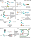
Multifaceted functions of lncRNAs in tumor and immune cells. (A) Two distinct functions of NKILA in tumor cells and T cells. In tumor cells, NKILA is upregulated by several inflammatory mediators (TNF-formula image and IL-1formula image) that inhibit the activation of NF-κB, which controls tumorigenesis and tumor progression. This regulation is blocked by miRNA targeting. In T cells, STAT1-mediated expression of NKILA inhibits NF-κB activity, thereby increasing immune cell death. Thus, the silencing of NKILA in tumor-reactive T cells enhances therapeutic effects in cancer by decreasing immune cell death. (B) Two distinct functions of NEAT1 in tumor cells and DCs. (C) Two distinct functions of NRON in tumor cells and T cells. (D) Two distinct functions of LUCAT1 in tumor cells and macrophages.
Figure 6
Figure 6
Detection of lncRNA markers via scRNA-seq versus bulk RNA-seq. The general procedure for scRNA-seq is summarized in the top panel. In the bottom panels, marker analyses at the bulk and single-cell levels are compared. Bulk RNA-seq mainly captures lncRNA markers that are differentially expressed in bulk tumor samples regardless of the cell type composition. scRNA-seq captures the lncRNA markers specific to certain cell types as well as markers specific to tumor-specific cell types.
Figure 7
Figure 7
Comparison of expressed lncRNAs and DEGs in scRNA-seq datasets versus bulk RNA-seq datasets. (A) TPM distribution for genes represented in single-cell RNA-seq datasets that were detected at ≥1 TPM in bulk RNA-seq data from TCGA (lung adenocarcinoma and squamous cell carcinoma for 10X and lung adenocarcinoma for SS2) in each scRNA-seq platform. (B) The proportion of single-cell DEG lncRNAs between tumor and nontumor samples overlapped with those from bulk RNA-seq data or pseudo-bulk RNA-seq data. Single-cell DEGs were acquired by comparing gene expression between single-cells from tumor and nontumor samples for each cell type. Bulk and pseudo-bulk DEGs were acquired by comparing gene expression between bulk RNA-seq data from paired tumor and nontumor samples and between pseudo-bulk data transformed from scRNA-seq data from tumor and nontumor tissue, respectively.

References

    1. Xu J, Bai J, Zhang X, et al. . A comprehensive overview of lncRNA annotation resources. Brief Bioinform 2017;18(2):236–49. - PubMed
    1. You B-H, Yoon S-H, Nam J-W. High-confidence coding and noncoding transcriptome maps. Genome Res 2017;27(6):1050–62. - PMC - PubMed
    1. Uszczynska-Ratajczak B, Lagarde J, Frankish A, et al. . Towards a complete map of the human long non-coding RNA transcriptome. Nat Rev Genet 2018;19(9):535–48. - PMC - PubMed
    1. Iyer MK, Niknafs YS, Malik R, et al. . The landscape of long noncoding RNAs in the human transcriptome. Nat Genet 2015;47(3):199–208. - PMC - PubMed
    1. Hon CC, Ramilowski JA, Harshbarger J, et al. . An atlas of human long non-coding RNAs with accurate 5' ends. Nature 2017;543(7644):199–204. - PMC - PubMed

Publication types

Substances