

Predicting RF Heating of Conductive Leads During Magnetic Resonance Imaging at 1.5 T: A Machine Learning Approach
- PMID: 34892151
- PMCID: PMC9940641
- DOI: 10.1109/EMBC46164.2021.9630718
Predicting RF Heating of Conductive Leads During Magnetic Resonance Imaging at 1.5 T: A Machine Learning Approach
Abstract
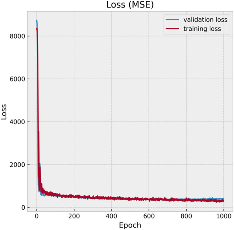
The number of patients with active implantable medical devices continues to rise in the United States and around the world. It is estimated that 50-75% of patients with conductive implants will need magnetic resonance imaging (MRI) in their lifetime. A major risk of performing MRI in patients with elongated conductive implants is the radiofrequency (RF) heating of the tissue surrounding the implant's tip due to the antenna effect. Currently, applying full-wave electromagnetic simulation is the standard way to predict the interaction of MRI RF fields with the human body in the presence of conductive implants; however, these simulations are notoriously extensive in terms of memory requirement and computational time. Here we present a proof-of-concept simulation study to demonstrate the feasibility of applying machine learning to predict MRI-induced power deposition in the tissue surrounding conductive wires. We generated 600 clinically relevant trajectories of leads as observed in patients with cardiac conductive implants and trained a feedforward neural network to predict the 1g-averaged SAR at the lead tips knowing only the background field of MRI RF coil and coordinates of points along the lead trajectory. Training of the network was completed in 11.54 seconds and predictions were made within a second with R2 = 0.87 and Root Mean Squared Error (RMSE) = 14.5 W/kg. Our results suggest that machine learning could provide a promising approach for safety assessment of MRI in patients with conductive leads.Clinical Relevance- Machine learning can potentially allow real-time assessment of MRI RF safety in patients with conductive leads when only the knowledge of lead's trajectory (image-based) and MRI RF coil features (vendor-specific) are in hand.
Figures


References
-
- Bhusal B et al., "Effect of Device Configuration and Patient's Body Composition on the RF Heating and Nonsusceptibility Artifact of Deep Brain Stimulation Implants During MRI at 1.5T and 3T," (in eng), J Magn Reson Imaging, vol. 53, no. 2, pp. 599–610, Feb 2021, doi: 10.1002/jmri.27346. - DOI - PubMed
-
- Golestanirad L, Keil B, Angelone LM, Bonmassar G, Mareyam A, and Wald LL, "Feasibility of using linearly polarized rotating birdcage transmitters and close-fitting receive arrays in MRI to reduce SAR in the vicinity of deep brain simulation implants," (in eng), Magn Reson Med, vol. 77, no. 4, pp. 1701–1712, Apr 2017, doi: 10.1002/mrm.26220. - DOI - PMC - PubMed
Publication types
MeSH terms
Grants and funding
LinkOut - more resources
Full Text Sources
Miscellaneous