. 2021 Dec 10;40(1):390.
doi: 10.1186/s13046-021-02191-3.

The IRF2/CENP-N/AKT signaling axis promotes proliferation, cell cycling and apoptosis resistance in nasopharyngeal carcinoma cells by increasing aerobic glycolysis

Affiliations

The IRF2/CENP-N/AKT signaling axis promotes proliferation, cell cycling and apoptosis resistance in nasopharyngeal carcinoma cells by increasing aerobic glycolysis

Cheng-Lin Qi et al. J Exp Clin Cancer Res. .

Abstract

Background: Centromere protein N (CENP-N) has been reported to be highly expressed in malignancies, but its role and mechanism in nasopharyngeal carcinoma (NPC) are unknown.

Methods: Abnormal CENP-N expression from NPC microarrays of GEO database was analyzed. CENP-N expression level was confirmed in NPC tissues and cell lines. Stable CENP-N knockdown and overexpression NPC cell lines were established, and transcriptome sequencing after CENP-N knockdown was performed. In vitro and in vivo experiments were performed to test the impact of CENP-N knockdown in NPC cells. ChIP and dual luciferase reporter assays were used to verify the combination of IRF2 and CENP-N. Western blot analysis, cellular immunofluorescence, immunoprecipitation and GST pulldown assays were used to verify the combination of CENP-N and AKT.

Results: CENP-N was confirmed to be aberrantly highly expressed in NPC tissues and cell lines and to be associated with high 18F-FDG uptake in cancer nests and poor patient prognosis. Transcriptome sequencing after CENP-N knockdown revealed that genes with altered expression were enriched in pathways related to glucose metabolism, cell cycle regulation. CENP-N knockdown inhibited glucose metabolism, cell proliferation, cell cycling and promoted apoptosis. IRF2 is a transcription factor for CENP-N and directly promotes CENP-N expression in NPC cells. CENP-N affects the glucose metabolism, proliferation, cell cycling and apoptosis of NPC cells in vitro and in vivo through the AKT pathway. CENP-N formed a complex with AKT in NPC cells. Both an AKT inhibitor (MK-2206) and a LDHA inhibitor (GSK2837808A) blocked the effect of CENP-N overexpression on NPC cells by promoting aerobic glycolysis, proliferation, cell cycling and apoptosis resistance.

Conclusions: The IRF2/CENP-N/AKT axis promotes malignant biological behaviors in NPC cells by increasing aerobic glycolysis, and the IRF2/CENP-N/AKT signaling axis is expected to be a new target for NPC therapy.

Keywords: AKT; Aerobic glycolysis; CENP-N; Gglucose metabolism; IRF2; NPC.


Conflict of interest statement

None.

Figures

Fig. 1
Fig. 1
CENP-N expression in NPC tissues and cells. a Representative images from NPC and NPG tissues with F-actin and CENP-N immunofluorescence staining (200×). b The expression of CENP-N in NPC and NPG tissues was evaluated by Western blotting, and the results showed that CENP-N was highly expressed in NPC cells. c Representative images from NPC and NPG tissue samples with low or high CENP-N immunohistochemical staining scores (4× and 200×). d Statistical analysis of the immunohistochemical results showed that CENP-N was highly expressed in NPC. e Representative 18F-FDG PET-CT images and SUVmax of NPC samples from NPC patients with low or high CENP-N expression (all 200×). f Survival curve of patients stratified by CENP-N expression. g Expression of CENP-N in NPC and NP460 cell lines. The data are shown as the mean ± SD values. * p < 0.05, ** p < 0.01, ***p < 0.001
Fig. 2
Fig. 2
RNA-sequencing after downregulating CENP-N. a qRT-PCR was used to verify CENP-N knockdown in 5-8F and CNE-2Z cell lines. b Western blotting was used to verify CENP-N knockdown in 5-8F and CNE-2Z cells. c Heat map showing that the genes altered after CENP-N knockdown were involved in glycolysis, apoptosis, proliferation, and cell cycling. d Bubble plot of KEGG pathway analysis results showing the function of genes altered after CENP-N knockdown. e Bar plot of GO analysis results showing the function of genes altered after CENP-N knockdown. f qRT-PCR was used to verify genes altered after CENP-N knockdown involved in cell glycolysis, apoptosis, proliferation, and cell cycle progression. g Western blotting was used to verify genes altered after CENP-N knockdown involved in glycolysis, apoptosis, proliferation, and cell cycling. The data are shown as the mean ± SD values. * p < 0.05, ** p < 0.01, ***p < 0.001
Fig. 3
Fig. 3
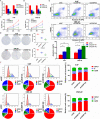
Changes in malignant biological behaviors of two NPC cell lines after downregulation of CENP-N. a Changes in relative cellular glucose uptake detected after knockdown of CENP-N in two NPC cell lines. b Changes in cellular lactate production detected after knockdown of CENP-N in two NPC cell lines. c Changes in cell viability detected by a CCK-8 assay after knockdown of CENP-N in two NPC cell lines. d Changes in cell proliferation detected after knockdown of CENP-N in two NPC cell lines. e Changes in the percentage of apoptotic cells detected after knockdown of CENP-N in two NPC cell lines. f Changes in the cell cycle distribution after knockdown of CENP-N in 5-8F and CNE-2Z cell lines. The data are shown as the mean ± SD values. * p < 0.05, ** p < 0.01, ***p < 0.001
Fig. 4
Fig. 4
Downregulation of CENP-N inhibited cell proliferation and glucose metabolism in vivo. a General view of tumor formation in nude mice. b The tumor volume curve of nude mice bearing tumors formed from 5-8F shCENP-N cells, CEN-2Z shCENP-N cells and the corresponding control cells. c Nude mice bearing tumors formed from 5-8F shCENP-N cells, CEN-2Z shCENP-N cells and the corresponding control cells. d The average tumor weight in nude mice bearing tumors formed from 5-8F shCENP-N cells, CEN-2Z shCENP-N cells and the corresponding control cells. e A representative PET-CT image was used to detect glucose uptake in 5-8F shCENP-N and CEN-2Z shCENP-N xenografts and the corresponding controls. f SUVmax values in nude mice bearing tumors. g SUVmean values in nude mice bearing tumors. h TLG values in nude mice bearing tumors. The data are shown as the mean ± SD values. * p < 0.05, ** p < 0.01
Fig. 5
Fig. 5
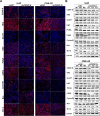
Expression levels of proteins related to cell proliferation, cell cycling, glucose metabolism and apoptosis were measured in xenografts formed from cells with CENP-N knockdown. a Immunofluorescence staining showed the expression of proteins related to cell proliferation, cell cycling, glycolysis and apoptosis in 5-8F shCENP-N and CEN-2Z shCENP-N xenografts and the corresponding controls (scale bar, 50 μm). b Western blot analysis showing the expression of proteins related to cell proliferation, cell cycling, glycolysis and apoptosis in 5-8F shCENP-N and CEN-2Z shCENP-N xenografts and the corresponding controls
Fig. 6
Fig. 6
CENP-N was regulated directly by IRF2 in NPC cells. a Sequence logo of the IRF2 binding site in the CENP-N promoter. b The GEPIA website was used to predict the correlation between IRF2 and CENP-N expression. c PCR analysis of the ChIP product showed that IRF2 bound to the CENP-N gene promoter region. d A luciferase reporter assay was used to detect the effects of IRF2 binding site in the promoter region of CENP-N. e Western blotting was used to verify the correlation between IRF2 and CENP-N expression. f Representative images from NPG and NPC tissue samples with low or high IRF2 immunohistochemical staining scores (4× and 200×). g Statistical analysis of the immunohistochemical results showed that IRF2 was highly expressed in NPC. The data are shown as the mean ± SD values. * p < 0.05, ** p < 0.01, ***p < 0.001
Fig. 7
Fig. 7
The tumor-promoting properties of CENP-N may be mediated via the AKT signaling pathway. a Changes in the expression of core proteins in the related signaling pathway after CENP-N knockdown in 5-8F and CNE-2Z cells. b Changes in AKT1, AKT2, and AKT3 mRNA expression after CENP-N knockdown in 5-8F and CNE-2Z cells. c An immunofluorescence assay was performed to detect the colocalization of CENP-N and AKT in 5-8F and CNE-2Z cells (scale bar, 10 μm). d The results of the coimmunoprecipitation assay demonstrated the interaction between CENP-N and AKT. e The results of the GST fusion protein pulldown assay demonstrated a direct interaction between CENP-N and AKT1. The data are shown as the mean ± SD values. ** p < 0.01, ***p < 0.001
Fig. 8
Fig. 8
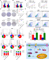
CENP-N promotes cell proliferation, cell cycling and apoptosis resistance by increasing aerobic glycolysis in NPC cells. a Changes in relative glucose uptake in two NPC cell lines were detected after CENP-N overexpression and GSK2837808A treatment. b Changes in lactic acid production in two NPC cell lines were detected after CENP-N overexpression and GSK2837808A treatment. c A CCK-8 assay was used to detect changes in cell viability after CENP-N overexpression and GSK2837808A treatment in two NPC cell lines. d Clone formation assay was used to detect the changes in cell proliferation after CENP-N overexpression and GSK2837808A treatment in two NPC cell lines. e The percentage of apoptotic cells was determined in two NPC cell lines after CENP-N overexpression and GSK2837808A treatment. f Changes in the cell cycle distribution in two NPC cell lines after CENP-N overexpression and GSK2837808A treatment. g Model showing the action mechanism of CENP-N in NPC. The data are shown as the mean ± SD values. * p < 0.05, ** p < 0.01, ***p < 0.001

References

    1. Johnson DE, Burtness B, Leemans CR, Lui VWY, Bauman JE, Grandis JR. Head and neck squamous cell carcinoma. Nat Rev Dis Primers. 2020;6(1):92. - PMC - PubMed
    1. Tang LL, Chen WQ, Xue WQ, He YQ, Zheng RS, Zeng YX, et al. Global trends in incidence and mortality of nasopharyngeal carcinoma. Cancer Lett. 2016;374(1):22–30. - PubMed
    1. Carioli G, Negri E, Kawakita D, Garavello W, La Vecchia C, Malvezzi M. Global trends in nasopharyngeal cancer mortality since 1970 and predictions for 2020: focus on low-risk areas. Int J Cancer. 2017;140(10):2256–2264. - PubMed
    1. Chen YP, Ismaila N, Chua MLK, Colevas AD, Haddad R, Huang SH, et al. Chemotherapy in combination with radiotherapy for definitive-intent treatment of stage II-IVA nasopharyngeal carcinoma: CSCO and ASCO guideline. J Clin Oncol. 2021;39(7):840–859. - PubMed
    1. Chittori S, Hong J, Saunders H, Feng H, Ghirlando R, Kelly AE, et al. Structural mechanisms of centromeric nucleosome recognition by the kinetochore protein CENP-N. Science. 2018;359(6373):339–343. - PMC - PubMed

Substances