. 2022 Feb;128(2):283-293.
doi: 10.1016/j.bja.2021.10.039. Epub 2021 Dec 8.

Reduction of NETosis by targeting CXCR1/2 reduces thrombosis, lung injury, and mortality in experimental human and murine sepsis

Affiliations

Reduction of NETosis by targeting CXCR1/2 reduces thrombosis, lung injury, and mortality in experimental human and murine sepsis

Mohmad Alsabani et al. Br J Anaesth. 2022 Feb.

Abstract

Background: Neutrophil extracellular traps (NETs) facilitate bacterial clearance but also promote thrombosis and organ injury in sepsis. We quantified ex vivo NET induction in septic humans and murine models of sepsis to identify signalling pathways that may be modulated to improve outcome in human sepsis.

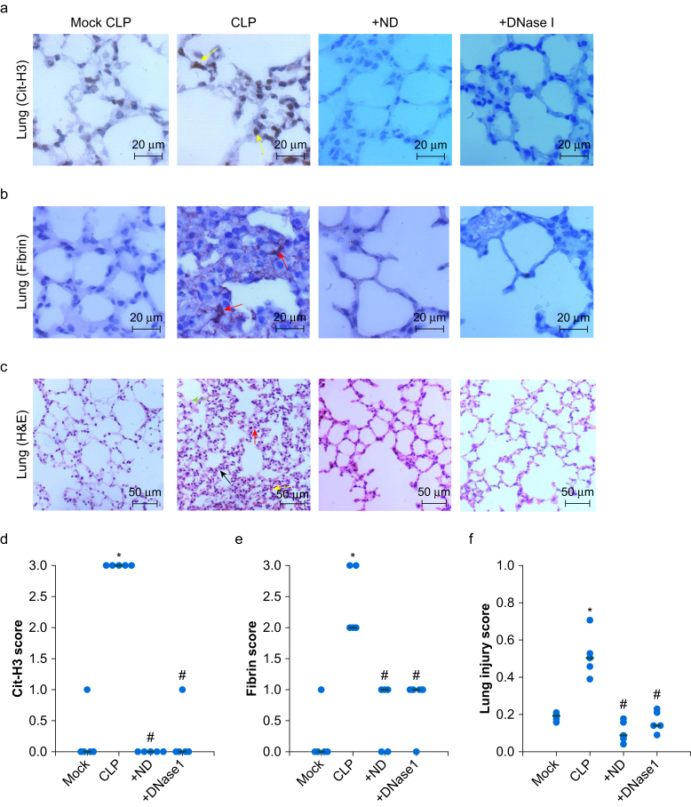
Methods: NET formation in human donor neutrophils was quantified after incubation with plasma obtained from patients with sepsis or systemic inflammation (double-blinded assessment of extracellular DNA using immunofluorescence microscopy). NET formation (% neutrophils forming NETs) was correlated with plasma cytokine levels (MultiPlex assay). Experimental sepsis (caecal ligation and puncture or intraperitoneal injection of Escherichia coli) was assessed in C57/BL6 male mice. The effect of pharmacological inhibition of CXCR1/2 signalling (reparixin) on NET formation, organ injury (hepatic, renal, and cardiac biomarkers), and survival in septic mice was examined.

Results: NET formation was higher after incubation with plasma from septic patients (median NETs=25% [10.5-46.5%]), compared with plasma obtained from patients with systemic inflammation (14% [4.0-23.3%]; P=0.02). Similar results were observed after incubation of plasma from mice with neutrophils from septic non-septic mice. Circulating CXCR1/2 ligands correlated with NETosis in patients (interleukin-8; r=0.643) and mice (macrophage inflammatory protein-2; r=0.902). In experimental sepsis, NETs were primarily observed in the lungs, correlating with fibrin deposition (r=0.702) and lung injury (r=0.692). Inhibition of CXCR1/2 using reparixin in septic mice reduced NET formation, multi-organ injury, and mortality, without impairing bacterial clearance.

Conclusion: CXCR1/2 signalling-induced NET formation is a therapeutic target in sepsis, which may be guided by ex vivo NET assays.

Keywords: CXCR1/2; interleukin-8; neutrophil depletion; neutrophil extracellular traps; organ injury; reparixin; sepsis.


Figures

Fig 1
Fig 1
Ex vivo neutrophil extracellular traps formation reflects in vivo NETosis. C57BL/6 male mice were anaesthetised and sepsis was induced either by caecal ligation and puncture (CLP) or intraperitoneal (i.p.) injection of Escherichia coli. Blood and tissues were collected at 4, 6, and 10 h after sepsis induction with mock CLP and saline (i.p.) as controls (three mice per time point per group). (a–d) Ex vivo NET formation assay with neutrophils isolated from normal mice and plasma from the mouse models and controls. Typical images of NET formation are presented in (a) and (b), where white arrows indicate normal neutrophils and yellow arrows indicate NETs. Scale bar=50 μm. NET formation was quantified as the percentage of neutrophils forming NETs per microscopic field and means (sd) are presented in (c) and (d). Mann–Whitney U-test, ∗P<0.05 increase NET formation compared with mock controls. (e, f) In vivo NET quantification. Representative images of immunohistochemically (IHC)-stained lung sections with an anti-citrullinated histone H3 (Cit-H3) antibody are presented (e, f). Arrows (yellow) indicate NETs. Scale bar=20 μm. Cit-H3 staining intensity was quantified and means (sd) are presented (g, h). Mann–Whitney U-test, ∗P<0.05 increase NET formation compared with mock controls. NETs, neutrophil extracellular traps; sd, standard deviation.
Fig 2
Fig 2
Neutrophil depletion reduces fibrin deposition and organ injury in sepsis. Typical images of lungs sections stained with anti-citrullinated histone H3 (Cit-H3) for neutrophil extracellular traps (NETs) (a) and anti-fibrin for fibrin deposition (b) (scale bar=20 μm). Yellow arrows indicate NETs and red arrows indicate fibrin deposition. (c) Lung sections were stained with haematoxylin and eosin (H&E). Typical images are presented. Red arrows indicate interstitial neutrophil, black arrows indicate membrane thickness, green arrows indicate proteinaceous debris, yellow arrows indicate hyaline membrane. Scale bar=50 μm. NET formation (d), fibrin score (e) and lung injury score (f) are presented. Six mice in each group. Mann–Whitney U-test, ∗P<0.05 compared with the mock group; #P<0.05 compared with the caecal ligation and puncture (CLP) group.
Fig 3
Fig 3
CXCL2–CXCR2 is important for neutrophil extracellular traps formation in sepsis. Circulating macrophage inflammatory protein-2 (MIP-2; a CXCL2 in mice) was measured in both caecal ligation and puncture (CLP) and Escherichia coli-induced sepsis models and controls (a, b) (n=3 per time point). Means (sd) are presented over the time course of the experiment. ∗P<0.05 increase in circulating MIP-2 compared with mock controls. (c) Representative fluorescence microscopy images showing the capability of phorbol 12-myristate 13-acetate (PMA) (positive control), interleukin (IL)-8 (100 pg ml−1) or MIP-2 (8000 pg ml−1)-spiked normal mouse plasma in inducing neutrophil extracellular traps (NETs) formation of isolated neutrophils from normal mice. Scale bar=50 μm. sd, standard deviation.
Fig 4
Fig 4
Reparixin, a CXCR2 inhibitor, reduces neutrophil extracellular traps formation in sepsis mice. Sepsis was induced in C57BL/6 male mice and treated with reparixin or saline as control (six mice per group). Blood and tissues were collected 10 h after sepsis induction. (a) Typical images of ex vivo neutrophil extracellular traps (NETs) formation are presented. (b) Typical images of anti-citrullinated histone H3 (Cit-H3) staining of lung sections for quantification of in vivo NETs formation are presented. White arrows indicate normal neutrophils; yellow arrows indicate NETs. Scale bar=50 μm. Quantification of ex vivo (c) and in vivo (d) NETs formation are presented. ∗Mann–Whitney U-test, P<0.05, compared sepsis model without (–inhibitor) to with reparixin treatment (+inhibitor). (e) Bacterial cultures of organ tissues, including heart, liver, spleen, lungs and kidneys at 10 h after cecal ligation and puncture (CLP)-sepsis induction. The means of colony-forming units (CFUs) from the organ tissues of septic mice treated with saline (–inhibitor) or with reparixin (+inhibitor) showed no difference.
Fig 5
Fig 5
Blocking CXCR2 reduces fibrin deposition, organ injury and improves survival times of sepsis mice. Typical photos of lungs 10 h after caecal ligation and puncture (CLP) and CLP treated with reparixin (CLP+inhibitor) (a) and typical images of haematoxylin and eosin (H&E) stained lung sections (b) are presented. Semi-quantified fibrin deposition in lung sections immunohistochemically (IHC)-stained with anti-fibrin antibody (c), lung injury scores (d), alanine aminotransferase (ALT) (e), blood urea nitrogen (BUN) (f) and cardiac troponin I (cTnI) (g) in mice 10 h after mock or CLP induction are presented. Mann–Whitney U test, ∗P<0.05 compared with mock group, #P<0.05 compared with the CLP group without treatment. (h) Kaplan–Meier survival curve is presented to compare CLP (n=8) with CLP treated with reparixin (n=6), log-rank test.

References

    1. Singer M., Deutschman C.S., Seymour C.W., et al. The third international consensus definitions for sepsis and septic shock (Sepsis-3) JAMA. 2016;315:801–810. - PMC - PubMed
    1. Rudd K.E., Johnson S.C., Agesa K.M., et al. Global, regional, and national sepsis incidence and mortality, 1990–2017: analysis for the Global Burden of Disease Study. Lancet. 2020;395:200–211. - PMC - PubMed
    1. Engelmann B., Massberg S. Thrombosis as an intravascular effector of innate immunity. Nat Rev Immunol. 2013;13:34–45. - PubMed
    1. Camicia G., Pozner R., de Larranaga G. Neutrophil extracellular traps in sepsis. Shock. 2014;42:286–294. - PubMed
    1. Brinkmann V., Reichard U., Goosmann C., et al. Neutrophil extracellular traps kill bacteria. Science. 2004;303:1532–1535. - PubMed

MeSH terms