. 2021 Dec 14;118(50):e2107389118.
doi: 10.1073/pnas.2107389118.

Biased M1 muscarinic receptor mutant mice show accelerated progression of prion neurodegenerative disease

Affiliations

Biased M1 muscarinic receptor mutant mice show accelerated progression of prion neurodegenerative disease

Miriam Scarpa et al. Proc Natl Acad Sci U S A. .

Abstract

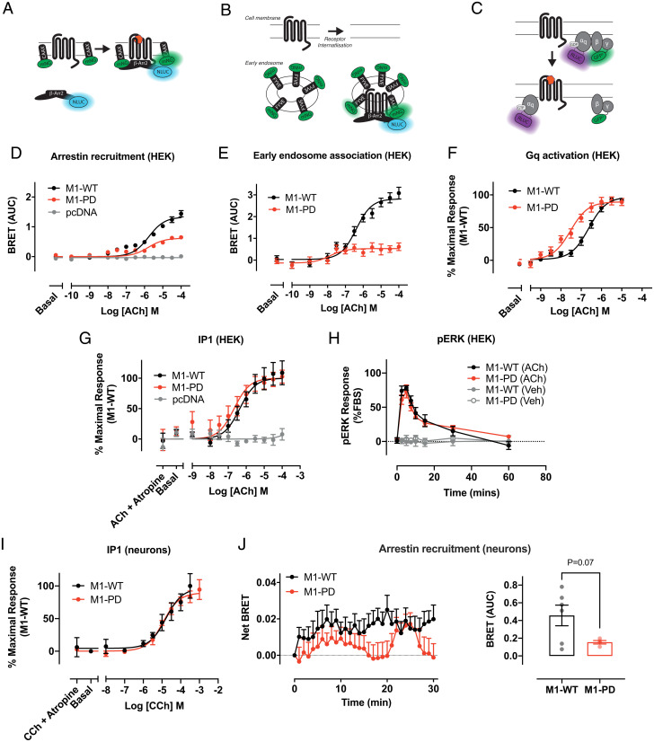
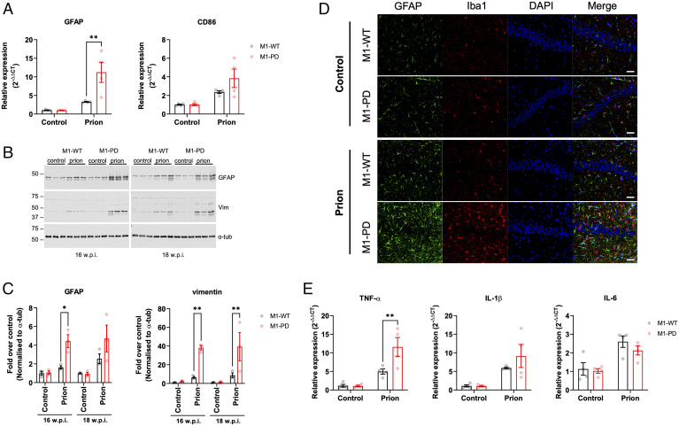
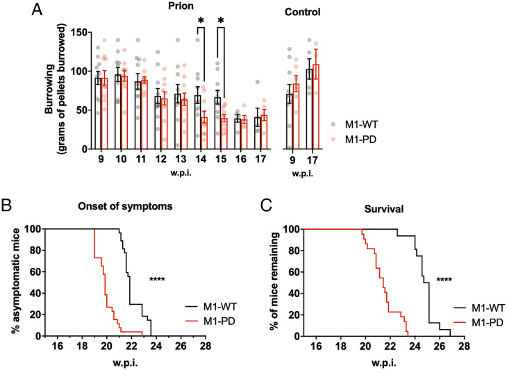
There are currently no treatments that can slow the progression of neurodegenerative diseases, such as Alzheimer's disease (AD). There is, however, a growing body of evidence that activation of the M1 muscarinic acetylcholine receptor (M1-receptor) can not only restore memory loss in AD patients but in preclinical animal models can also slow neurodegenerative disease progression. The generation of an effective medicine targeting the M1-receptor has however been severely hampered by associated cholinergic adverse responses. By using genetically engineered mouse models that express a G protein-biased M1-receptor, we recently established that M1-receptor mediated adverse responses can be minimized by ensuring activating ligands maintain receptor phosphorylation/arrestin-dependent signaling. Here, we use these same genetic models in concert with murine prion disease, a terminal neurodegenerative disease showing key hallmarks of AD, to establish that phosphorylation/arrestin-dependent signaling delivers neuroprotection that both extends normal animal behavior and prolongs the life span of prion-diseased mice. Our data point to an important neuroprotective property inherent to the M1-receptor and indicate that next generation M1-receptor ligands designed to drive receptor phosphorylation/arrestin-dependent signaling would potentially show low adverse responses while delivering neuroprotection that will slow disease progression.

Keywords: GPCR; M1 muscarinic acetylcholine receptor; neurodegenerative disease; phosphorylation.


Conflict of interest statement

The authors declare no competing interest.

Figures

Fig. 1.
Fig. 1.
Arrestin recruitment and receptor internalization of the M1-receptor are dependent on receptor phosphorylation. (A–C) Schematic of the bystander BRET assays for arrestin recruitment to the M1-receptor (A), receptor translocation to early endosomes (B), and Gq activation to the M1-receptor (C). (D) ACh-stimulated translocation of β-arrestin-2 to the cell membrane in HEK293T cells transfected with pcDNA3, M1-WT, or M1-PD assessed by bystander BRET. Data are expressed as mean ± SEM of five independent experiments performed in triplicates. (E) Translocation of M1-WT and M1-PD to early endosomes in response to ACh treatment assessed through a bystander BRET assay. Data are expressed as mean ± SEM of five independent experiments performed in triplicates. (F) ACh-stimulated Gαq activation by the M1-WT or M1-PD receptor measured by a decrease in BRET. Data are expressed as means ± SEM of four independent experiments performed in quadruplicate. (G) IP1 accumulation after 60-min stimulation with ACh in HEK cells transiently transfected with the M1-WT and M1-PD constructs or the empty vector (pcDNA). Data are expressed as means ± SEM of four to seven independent experiments performed in duplicate or quadruplicate. (H) Time course of pERK signaling in HEK293T cells transfected with M1-WT or M1-PD and stimulated with ACh (100 μM) or vehicle (0.01% dimethyl sulfoxide). Data are expressed as means ± SEM of three independent experiments performed in triplicate (n = 3). (I) IP1 accumulation after 60-min stimulation with CCh in primary hippocampal-cortical neurons prepared from M1-WT or M1-PD mice. Data are expressed as means ± SEM of three independent experiments performed in duplicate. (J) Time course (Left) of ACh (100 μM)-stimulated translocation of β-arrestin-2 to the cell membrane in primary hippocampal-cortical neurons prepared from M1-WT or M1-PD mice. Mean area under the curve (AUC) is shown on the Right. Data are expressed as mean ± SEM of four to six independent experiments performed in triplicates.
Fig. 2.
Fig. 2.
Prion-infected M1-PD mice show accelerated appearance of disease markers in the hippocampus compared to M1-WT mice. Lysates were prepared from the hippocampus of control or prion-infected M1-WT and M1-PD mice at 16 and 18 w.p.i., and Western blot analysis was used to analyze the expression of a panel of pathological markers. (A) Lysates were incubated in the presence or absence of proteinase K prior to Western blot to detect nondigested scrapie prion protein (PrPsc) and total prion protein (PrPtot), respectively. Band analysis for PrPsc and PrPtot expression in (B) is shown as means ± SEM of a ratio of α-tubulin expression. (C) APO-E, serpinA3N, clusterin, and galectin-1 were detected in the hippocampus and band analysis is shown in D as means ± SEM of a ratio of α-tubulin expression relative to control-infected M1-WT (n = 3 mice). All data were analyzed using two-way ANOVA with Sidak multiple comparisons where *P < 0.05, **P < 0.01, ***P < 0.001 (M1-WT versus M1-PD).
Fig. 3.
Fig. 3.
Neuroinflammation is exacerbated in the hippocampus of prion-infected M1-PD mice compared to M1-WT controls. (A) mRNA levels of GFAP and CD86, markers of astrocytes and microglia, respectively, were quantified using quantitative RT-PCR of hippocampus from control or prion-diseased M1-WT or M1-PD mice at 16 w.p.i. Data are expressed as means ± SEM of a ratio of α-tubulin RNA expression relative to M1-WT (n = 4 mice). **P < 0.01, two-way ANOVA with Sidak multiple comparisons (M1-WT versus M1-PD). (B and C) Astrogliosis in the hippocampus was assessed using Western blot analysis of lysates prepared from control or prion-infected mice at 16 and 18 w.p.i. Lysates were probed for astrocytic markers GFAP and vimentin (vim), and α-tubulin (α-tub) antibody was used as a loading control. (C) Band analysis for each blot was performed, and data are shown as means ± SEM of a ratio of α-tubulin relative to control M1-WT (n = 3 mice). *P < 0.05, **P < 0.01, two-way ANOVA Sidak multiple comparisons (M1-WT versus M1-PD). (D) Immunohistochemical staining for GFAP and Iba-1 in the hippocampus of control and prion-infected M1-WT and M1-PD mice at 16 w.p.i. The nuclei were stained blue with DAPI. (Scale bar, 100 μm.) (E) Quantitative RT-PCR showing the expression of proinflammatory (TNF-α, IL-1β, IL-6) cytokines in the hippocampus of control and prion-infected M1-WT and M1-PD mice at 16 w.p.i. Data are expressed as a ratio of α-tubulin RNA expression relative to control M1-WT (n = 4 mice). Data were analyzed using two-way ANOVA with Sidak’s multiple comparisons, where **P < 0.01 (M1-WT versus M1-PD).
Fig. 4.
Fig. 4.
Removal of M1-receptor phosphorylation sites accelerates prion disease and decreases survival time. (A) Burrowing responses (food pellets [grams] displaced from the tube) of control or prion-infected M1-WT and M1-PD mice were assessed from 9 w.p.i. (n = 3 to 10 mice; *P < 0.05; two-way ANOVA or mixed-effects model with uncorrected Fisher’s least significant difference test). Onset of at least two early indicators of prion disease (n = 26 to 27) (B) and Kaplan–Meier survival plot (n = 16 to 22) (C) for prion-infected M1-WT and M1-PD. Curves were analyzed with a Gehan–Breslow–Wilcoxon test, where ****P < 0.0001.

References

    1. Prince M., et al. , “World Alzheimer Report 2015: The global impact of dementia” (Alzheimer’s Disease International, London, 2015).
    1. Courtney C., et al. ; AD2000 Collaborative Group, Long-term donepezil treatment in 565 patients with Alzheimer’s disease (AD2000): Randomised double-blind trial. Lancet 363, 2105–2115 (2004). - PubMed
    1. Inglis F., The tolerability and safety of cholinesterase inhibitors in the treatment of dementia. Int. J. Clin. Pract. Suppl. 127, 45–63 (2002). - PubMed
    1. Svensson A.-L., Alafuzoff I., Nordberg A., Characterization of muscarinic receptor subtypes in Alzheimer and control brain cortices by selective muscarinic antagonists. Brain Res. 596, 142–148 (1992). - PubMed
    1. Levey A. I., Immunological localization of m1-m5 muscarinic acetylcholine receptors in peripheral tissues and brain. Life Sci. 52, 441–448 (1993). - PubMed

Publication types

MeSH terms

Substances

LinkOut - more resources