Review
. 2021 Dec 10;4(1):1387.
doi: 10.1038/s42003-021-02910-8.

Towards organoid culture without Matrigel

Affiliations
Review

Towards organoid culture without Matrigel

Mark T Kozlowski et al. Commun Biol. .

Abstract

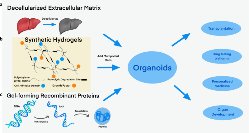
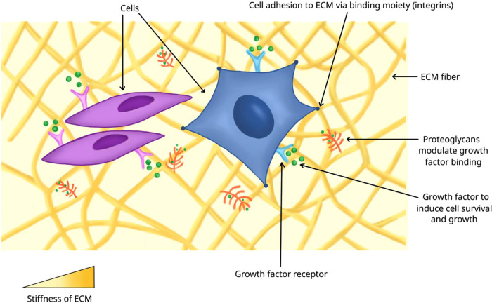
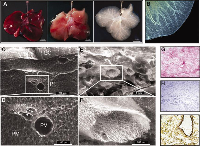
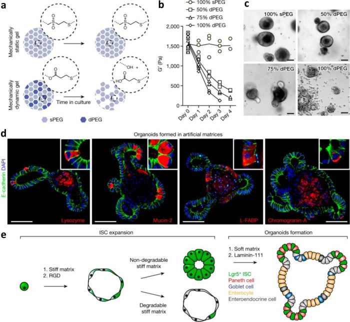
Organoids-cellular aggregates derived from stem or progenitor cells that recapitulate organ function in miniature-are of growing interest in developmental biology and medicine. Organoids have been developed for organs and tissues such as the liver, gut, brain, and pancreas; they are used as organ surrogates to study a wide range of questions in basic and developmental biology, genetic disorders, and therapies. However, many organoids reported to date have been cultured in Matrigel, which is prepared from the secretion of Engelbreth-Holm-Swarm mouse sarcoma cells; Matrigel is complex and poorly defined. This complexity makes it difficult to elucidate Matrigel-specific factors governing organoid development. In this review, we discuss promising Matrigel-free methods for the generation and maintenance of organoids that use decellularized extracellular matrix (ECM), synthetic hydrogels, or gel-forming recombinant proteins.


Conflict of interest statement

The authors declare no competing interests.

Figures

Fig. 1
Fig. 1. Methods of making organoids without Matrigel.
Replacing the undefined medium of Matrigel is a major goal of organoid culture. We will discuss three main alternative media: (a) decellularized extracellular matrix and other derived proteins, (b) synthetic hydrogels, which generally incorporate cell-adhesive domains or proteolytic degradation sites, and (c) gel-forming recombinant peptides. Adding multipotent cells to these matrices enables the growth of organoids, which are potentially applicable as transplants, drug-testing platforms, personalized medicine, and means to understand organ development.
Fig. 2
Fig. 2. Microenvironment of cells.
Cells in an organ or organoid are surrounded by other cells, extracellular matrix (ECM) proteins, and growth factors sequestered in the proteoglycan-modified ECM proteins. Cells bind to ECM proteins via adhesion molecules, such as integrin receptors, which provide signaling cues to exert biological functions. The stiffness of ECM experienced by the cells also affects their biological activities.
Fig. 3
Fig. 3. An example of whole-organ ferret liver decellularization with excellent retention of structural information, for use as an organoid scaffold.
Figure (a) shows the liver at the start of treatment, then after 20 and 120 min of decellularization. A micrograph of the decellularized liver, (b), shows that the liver capsule and vasculature remain intact after cell removal. Scanning electron microscope images show that the structure of the liver is remarkably well-conserved, with an intact Glisson’s capsule (GC) visible in (c), and an intact hepatic artery (HA), hepatic portal vein (PV), and biliary duct (BD) visible in (d). Other blood vessels are structurally intact, despite cell removal (e), with the structural details apparent even at high magnification (f). H&E staining (g) shows that all cells have been removed, with the pink stain showing protein-containing extracellular matrix; this absence of cellular material is further confirmed by Mason’s Trichrome staining (h). Movat-Pentachrome staining (i) shows the presence of collagen in yellow, and a dark stain shows elastin around an artery. Decellularization can proceed gently enough to retain structural information, yielding scaffolds that can be colonized by pluripotent cells which then differentiate into mature organoids. The image is from ref. and is reproduced with permission.
Fig. 4
Fig. 4. Growth of intestinal organoids on synthetic hydrogels, and effects of matrix stiffness and degradability on their formation.
Gjorevski and colleagues demonstrated defined PEG-based intestinal organoid culture. The stiffness and degradability has a major effect on the ability of induced pluripotent cells to differentiate into intestinal organoids. By varying the ratio of hydrolytically labile functional groups (dPEG) to stable functional groups (sPEG), the rate of degradation of the gel can be controlled (a, b). Higher ratios of sPEG are associated with the expansion of intestinal stem cells, whereas the degradable gels lead to the formation of organoids containing differentiated cells (c). In fact, organoid formation is observed only in gels that have a stiffness of ~190 Pa: cells expressing lysozyme (Paneth cells), mucin-2 (Goblet cells), and Chromogranin-A (enteroendocrine cells) are in different compartments, indicating that specialized cells are spatially separated (d). In short, a stiff matrix leads to intestinal stem cell proliferation and expansion, but a soft matrix and functionalization with laminin-111 promotes differentiation (e). The image is from ref. and is reproduced with permission.
Fig. 5
Fig. 5. Growth of cardiomyocytes on recombinant proteins, and effects of elastic modulus on cardiomyocyte differentiation.
The elastin-like proteins (ELPs) used by Chung and colleagues (a) consist of a bioactive domain translationally fused to one or more elastin-like domains; these domains contain lysine groups to facilitate crosslinking by tetrakis hydroxymethyl phosphonium chloride (THPC). By varying the ratio of THPC to ELP reactive groups, it is possible to tune the elastic modulus of the resulting culture matrix (b) without significantly altering the diffusion of nutrients or other vital factors through the gel (c). Embryoid bodies embedded in the matrix undergo differentiation into cardiomyocytes most favorably in the gels with the lowest elastic modulus (d); the cells show the greatest contractility when grown in protein crosslinked with a 0.5:1 ratio of THPC:ELP reactive groups. The image is from ref. and is reproduced with permission.
Fig. 6
Fig. 6. Generating pancreatic organoids with a recombinant ECM protein.
An artificial elastin-like polypeptide functionalized with a sequence from laminin can be used to generate organoids from pancreatic ductal progenitor cells from adult mice. (a) The recombinant protein (named aECM-lam) incorporates an IKVAV-containing 18-amino acid sequence derived from α1 laminin. The aECM-scr is a scrambled sequence control for aECM-lam. (b) aECM-lam permits the differentiation of endocrine (expressing C-peptide and glucagon) and acinar cell lineages (expressing amylase). (c) Individual organoids (Endocrine/Acinar) grown in aECM-lam express beta-cell maturation markers glucokinase, Pcsk1, and Pcsk2. (d) Organoids grown in aECM-lam are capable of secreting insulin in vitro when challenged by high concentrations of d-glucose or a combination of d-glucose and cAMP activator theophylline. The image is from ref. and is reproduced with permission.

References

    1. Lancaster MA, Knoblich JA. Organogenesis in a dish: modeling development and disease using organoid technologies. Science. 2014;345:10. - PubMed
    1. Grapin-Botton A. Three-dimensional pancreas organogenesis models. Diabetes Obes. Metab. 2016;18:33–40. - PMC - PubMed
    1. Boj SF, et al. Organoid models of human and mouse ductal pancreatic cancer. Cell. 2015;160:324–338. - PMC - PubMed
    1. Qian XY, Nguyen HN, Jacob F, Song HJ, Ming GL. Using brain organoids to understand Zika virus-induced microcephaly. Development. 2017;144:952–957. - PMC - PubMed
    1. Choi H, Song J, Park G, Kim J. Modeling of autism using organoid technology. Mol. Neurobiol. 2017;54:7789–7795. - PubMed

Publication types