. 2022 Jan;21(1):e13521.
doi: 10.1111/acel.13521. Epub 2021 Dec 10.

Loss of lamin-B1 and defective nuclear morphology are hallmarks of astrocyte senescence in vitro and in the aging human hippocampus

Affiliations

Loss of lamin-B1 and defective nuclear morphology are hallmarks of astrocyte senescence in vitro and in the aging human hippocampus

Isadora Matias et al. Aging Cell. 2022 Jan.

Abstract

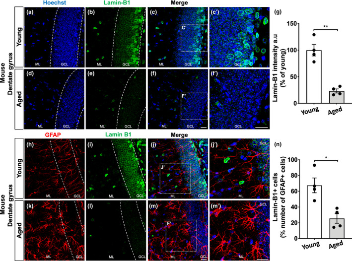
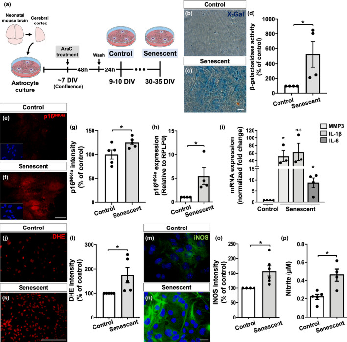
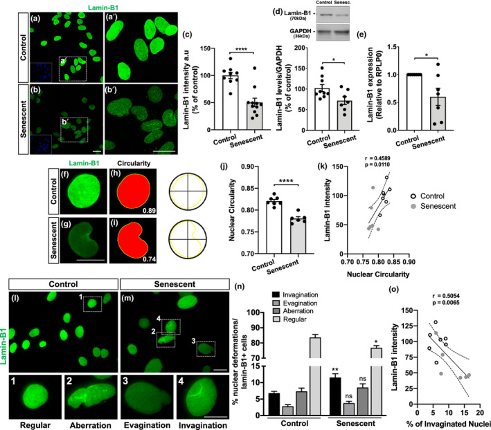
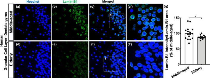
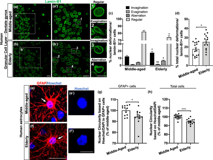
The increase in senescent cells in tissues, including the brain, is a general feature of normal aging and age-related pathologies. Senescent cells exhibit a specific phenotype, which includes an altered nuclear morphology and transcriptomic changes. Astrocytes undergo senescence in vitro and in age-associated neurodegenerative diseases, but little is known about whether this process also occurs in physiological aging, as well as its functional implication. Here, we investigated astrocyte senescence in vitro, in old mouse brains, and in post-mortem human brain tissue of elderly. We identified a significant loss of lamin-B1, a major component of the nuclear lamina, as a hallmark of senescent astrocytes. We showed a severe reduction of lamin-B1 in the dentate gyrus of aged mice, including in hippocampal astrocytes, and in the granular cell layer of the hippocampus of post-mortem human tissue from non-demented elderly. The lamin-B1 reduction was associated with nuclear deformations, represented by an increased incidence of invaginated nuclei and loss of nuclear circularity in senescent astrocytes in vitro and in the aging human hippocampus. We also found differences in lamin-B1 levels and astrocyte nuclear morphology between the granular cell layer and polymorphic layer in the elderly human hippocampus, suggesting an intra-regional-dependent aging response of human astrocytes. Moreover, we described senescence-associated impaired neuritogenic and synaptogenic capacity of mouse astrocytes. Our findings show that reduction of lamin-B1 is a conserved feature of hippocampal cells aging, including astrocytes, and shed light on significant defects in nuclear lamina structure which may contribute to astrocyte dysfunctions during aging.

Keywords: aging; astrocyte; human and mouse hippocampus; lamin-B1; senescence; synapse.


Conflict of interest statement

The authors declare no conflict of interest.

Figures

FIGURE 1
FIGURE 1
Age‐related loss of lamin‐B1 in the mouse hippocampus. (a–g) Densitometric analysis of lamin‐B1 staining in the mouse hippocampal dentate gyrus, including the molecular layer (ML) and granular cell layer (GCL), revealed a global reduction of lamin‐B1 intensity in aged mice compared with young mice (p = 0.0033). (h–n) Decreased proportion of lamin‐B1 + cells in relation to the total number of GFAP + cells in the dentate gyrus of aged mice compared with young ones (p = 0.0126). Significance was determined using unpaired t test with Welch's correction. Error bars represent ±SEM. Individual data points are plotted and represent individual animals (n = 4 animals per experimental group). Scale bars, 20 µm
FIGURE 2
FIGURE 2
Characterization of a new in vitro model for astrocyte senescence. (a) Primary murine astrocyte cultures were maintained for 9–10 DIV (control group) or 30–35 DIV (senescent group) and analyzed for several senescence‐associated biomarkers. (b–d) Astrocytes cultured for 30–35 DIV showed increased SA‐β‐galactosidase activity compared with control cultures (p = 0.0481). (e–g) Higher immunostaining for p16INK4a in senescent astrocytes compared with control cultures (p = 0.0459). (h) Senescent astrocytes showed increased expression of p16INK4a (p = 0.0471). (i) Expression levels of MMP3 and IL‐6 were increased in senescent astrocyte cultures compared with control ones (p = 0.0267 and p = 0.0142, respectively). (j–l) Increased intensity of DHE labeling (level of ROS) in senescent astrocytes compared with control cultures (p = 0.0467). (m–o) Higher immunostaining for iNOS in senescent astrocytes (p = 0.0311). (p) Nitrite production was elevated in senescent astrocytes (p = 0.0100). Significance was determined using unpaired t test. Error bars represent ±SEM. Individual data points are plotted and represent individual cultures (n = 3–6 cultures per experimental group). Scale bars, 50 µm in (c) and 20 µm in (f), (k), and (n)
FIGURE 3
FIGURE 3
Senescent astrocytes present impaired neuritogenic and synaptogenic properties in vitro. (a) Neural progenitor cell cultures were maintained in DMEM (vehicle), ACM‐Control, or ACM‐Senescent for 2 DIV. (b–d) The number of primary neurites per cell was quantified based on β‐Tubulin III immunostaining. (e) Neural progenitor cells treated with ACM‐Control showed increased number of neurites compared with those treated with DMEM (p = 0.0097). In contrast, neural progenitors treated with ACM‐Senescent exhibited reduced neurite number compared with ACM‐Control (p = 0.0044). (a, f–h) Mature neurons (12 DIV) were treated with DMEM, ACM‐Control, or ACM‐Senescent for 3 h, and the number of synapses was quantified based on synaptophysin/spinophilin colocalization puncta. (i) Neurons treated with ACM‐Control showed increased percentage of colocalized puncta compared with those treated with DMEM (p = 0.0083). Conversely, ACM‐Senescent reduced the number of synapses compared with ACM‐Control (0.0010). Significance was determined using one‐way ANOVA with Tukey's multiple comparisons test. Error bars represent ±SEM. Individual data points are plotted and represent individual cultures (n = 3–9 cultures per experimental condition). Scale bars, 20 µm
FIGURE 4
FIGURE 4
Nuclear deformations are associated with lamin‐B1 loss in senescent astrocytes. (a–c) Reduced immunostaining for lamin‐B1 in senescent astrocytes compared with the control group (p < 0.0001). (d) Lamin‐B1 protein levels were diminished in senescent astrocytes in comparison with the control group (p = 0.0329). (e) Senescent astrocytes showed decreased expression of lamin‐B1 (p = 0.0417). (f–i) Nuclear circularity analysis is based on the area and perimeter of the nucleus. Circularity has a maximum value of 1 and diminishes as the nuclear shape becomes increasingly convoluted, as observed in senescent cells (g, i). (j) Senescent astrocytes displayed reduced nuclear circularity compared with the control group (p < 0.0001). (k) A positive correlation was observed between lamin‐B1 intensity and the nuclear circularity value (r = 0.4589; p = 0.0110). (l, m) Distinct nuclear morphological profiles were evaluated, such as regular (1), aberration (2), evagination (3), and invagination (4) in control and senescent cultured astrocytes, based on lamin‐B1 staining. (n) Senescent astrocyte showed an increased incidence of invaginated nuclei (p = 0.0054) and a decreased proportion of regular nuclei (p = 0.0263). (o) A negative correlation was observed between lamin‐B1 intensity and the incidence of invaginated nuclei (r = 0.5054; p = 0.0065). Significance was determined using unpaired t test with Welch's correction. Linear regression opting to show 95% confidence bands of the best‐fit line. Error bars represent ±SEM. Individual data points are plotted and represent individual cultures (n = 6–11 cultures per experimental condition). Control cultures are represented by white dots and senescent cultures by gray dots in (k) and (o). Scale bars, 20 µm in (b), (b′) and (M); 10 µm in (g) and (4)
FIGURE 5
FIGURE 5
Lamin‐B1 reduction in the human dentate gyrus upon aging. (a–g) Densitometric analysis of lamin‐B1 staining at the hippocampal granular cell layer from post‐mortem human tissue revealed an overall reduction of lamin‐B1 intensity in elderly cases compared with middle‐aged ones (p = 0.0308). n = 16 and 13 individuals for middle‐aged and elderly groups, respectively. Scale bars, 20 µm. Significance was determined using unpaired t test with Welch's correction. Error bars represent ±SEM. Individual data points are plotted and represent individual donors
FIGURE 6
FIGURE 6
Neural cells, including astrocytes, from the granular cell layer of the human hippocampus undergo nuclear abnormalities upon aging. (a, b) Distinct nuclear morphological profiles were evaluated, such as regular (1), evagination (2), invagination (3), and aberration (4) at the hippocampal granular cell layer in post‐mortem human tissue from middle‐aged and elderly donors based on lamin‐B1 staining. Scale bars, 20 µm in (b) and (b′); 10 µm in (4). (c) Elderly donors exhibited a higher incidence of invaginated (p = 0.0494) and aberrant nuclei (p = 0.0243), resulting in a decreased proportion of regular nuclei (p = 0.0102), compared with the middle‐aged group. n = 16 and 14 individuals for middle‐aged and elderly groups, respectively. (d) Elderly donors showed an increased proportion of total nuclear deformations (ie, evagination + invagination + aberration) (p = 0.0119). n = 16 and 14 individuals for middle‐aged and elderly groups, respectively. (e–g) Nuclear circularity was quantified based on Hoechst or DAPI staining in post‐mortem human tissue from middle‐aged (e–e′) and elderly donors (f–f′). Elderly donors presented a reduced nuclear circularity based on the number of GFAP+cells (g; p = 0.0243; n = 12 individuals for both age groups) and on the total number of cells (h; p < 0.0001; n = 15 and 13 individuals for middle‐aged and elderly groups, respectively). Scale bars, 20 µm in (f) and 10 µm in (f′). Significance was determined using unpaired t test with Welch's correction. Error bars represent ± SEM. Individual data points are plotted and represent individual donors

References

    1. Baldwin, K. T. , & Eroglu, C. (2017). Molecular mechanisms of astrocyte‐induced synaptogenesis. Current Opinion in Neurobiology, 45, 113–120. 10.1016/j.conb.2017.05.006 - DOI - PMC - PubMed
    1. Barbar, L. , Jain, T. , Zimmer, M. , Kruglikov, I. , Sadick, J. S. , Wang, M. , Kalpana, K. , Rose, I. V. L. , Burstein, S. R. , Rusielewicz, T. , Nijsure, M. , Guttenplan, K. A. , di Domenico, A. , Croft, G. , Zhang, B. , Nobuta, H. , Hébert, J. M. , Liddelow, S. A. , & Fossati, V. (2020). CD49f is a novel marker of functional and reactive human iPSC‐derived astrocytes. Neuron, 107(3), 436–453.e12. 10.1016/j.neuron.2020.05.014 - DOI - PMC - PubMed
    1. Bedrosian, T. A. , Houtman, J. , Eguiguren, J. S. , Ghassemzadeh, S. , Rund, N. , Novaresi, N. M. , Hu, L. , Parylak, S. L. , Denli, A. M. , Randolph‐Moore, L. , Namba, T. , Gage, F. H. , & Toda, T. (2020). Lamin B1 decline underlies age‐related loss of adult hippocampal neurogenesis. EMBO Journal, 40(3), e105819. 10.15252/embj.2020105819 - DOI - PMC - PubMed
    1. Bellaver, B. , Souza, D. G. , Souza, D. O. , & Quincozes‐Santos, A. (2017). Hippocampal astrocyte cultures from adult and aged rats reproduce changes in glial functionality observed in the aging brain. Molecular Neurobiology, 54, 2969–2985 10.1007/s12035-016-9880-8 - DOI - PubMed
    1. Bhat, R. , Crowe, E. P. , Bitto, A. , Moh, M. , Katsetos, C. D. , Garcia, F. U. , Johnson, F. B. , Trojanowski, J. Q. , Sell, C. , & Torres, C. (2012). Astrocyte senescence as a component of Alzheimer’s disease. PLoS One, 7, e45069 10.1371/journal.pone.0045069 - DOI - PMC - PubMed

Publication types