. 2022 Feb;9(4):e2103098.
doi: 10.1002/advs.202103098. Epub 2021 Dec 11.

Hierarchically Multivalent Peptide-Nanoparticle Architectures: A Systematic Approach to Engineer Surface Adhesion

Affiliations

Hierarchically Multivalent Peptide-Nanoparticle Architectures: A Systematic Approach to Engineer Surface Adhesion

Woo-Jin Jeong et al. Adv Sci (Weinh). 2022 Feb.

Abstract

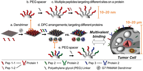
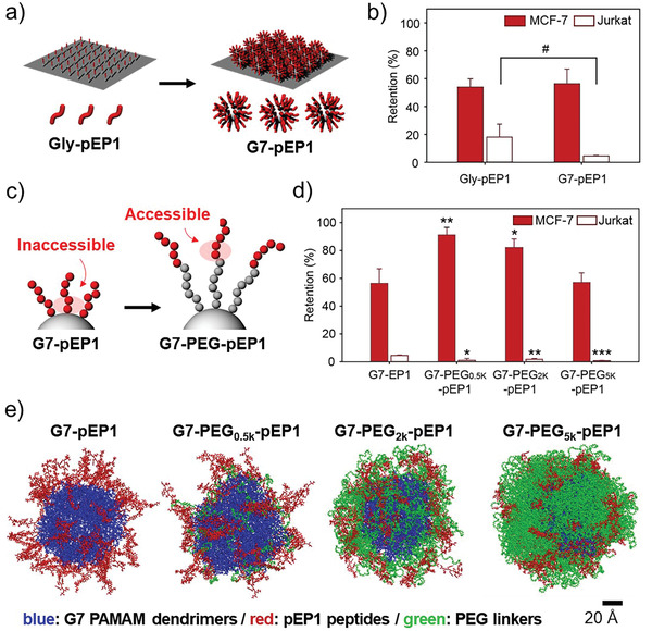
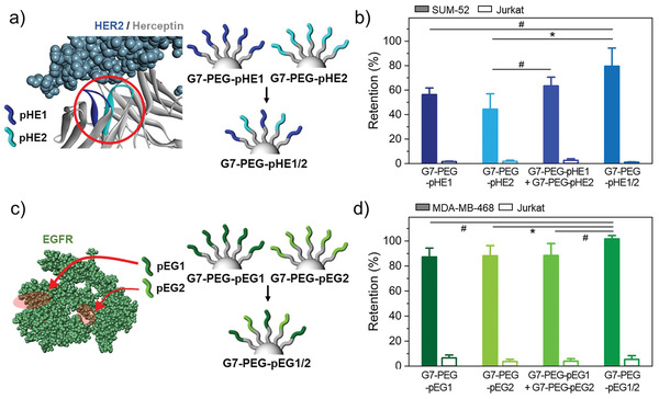
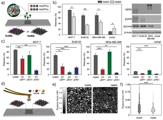
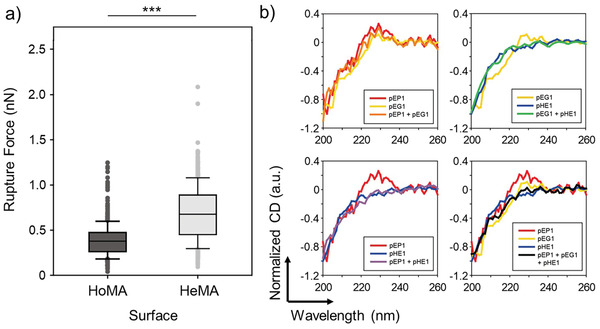
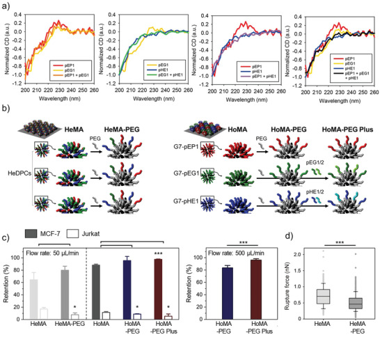
The multivalent binding effect has been the subject of extensive studies to modulate adhesion behaviors of various biological and engineered systems. However, precise control over the strong avidity-based binding remains a significant challenge. Here, a set of engineering strategies are developed and tested to systematically enhance the multivalent binding of peptides in a stepwise manner. Poly(amidoamine) (PAMAM) dendrimers are employed to increase local peptide densities on a substrate, resulting in hierarchically multivalent architectures (HMAs) that display multivalent dendrimer-peptide conjugates (DPCs) with various configurations. To control binding behaviors, effects of the three major components of the HMAs are investigated: i) poly(ethylene glycol) (PEG) linkers as spacers between conjugated peptides; ii) multiple peptides on the DPCs; and iii) various surface arrangements of HMAs (i.e., a mixture of DPCs each containing different peptides vs DPCs cofunctionalized with multiple peptides). The optimized HMA configuration enables significantly enhanced target cell binding with high selectivity compared to the control surfaces directly conjugated with peptides. The engineering approaches presented herein can be applied individually or in combination, providing guidelines for the effective utilization of biomolecular multivalent interactions using DPC-based HMAs.

Keywords: binding avidity; dendrimer-peptide conjugate; hierarchically multivalent architectures; multivalent binding; peptide engineering.


Conflict of interest statement

The authors declare no conflict of interest.

Figures

Figure 1
Figure 1
Schematic illustration of the stepwise engineering strategies applied to control the multivalent binding behaviors of various peptides with G7 PAMAM dendrimers: a) hyperbranched PAMAM dendrimers were employed to increase local peptide density; b) PEG spacers were introduced to spatially segregate the peptides from the dendrimer and to increase the conformational freedom of the peptides; c) multiple peptide sequences that bind to different sites within a single protein were coimmobilized to the dendrimer‐PEG surfaces to enhance the target binding; d) two different HMA surfaces, a mixture of DPCs each containing different peptides (HoMA) and DPCs cofunctionalized with multiple peptides (HeMA), were tested to investigate the effect of the peptide arrangement on their hetero‐multivalent binding behaviors; e) PEG linkers were introduced to hetero‐HMAs to explore whether the use of spacer molecules on HMA synergize the multivalent binding effect.
Figure 2
Figure 2
Surface engineering of individual DPCs: a) Introduction of G7 PAMAM dendrimers as a building block of HMA formation. b) % retention of surface‐bound MCF‐7 (EpCAMhigh; red) and Jurkat (EpCAMnegative; white) cells on Gly‐pEP1 versus G7‐pEP1 upon washing with a high shear flow. c) Schematic illustration of anticipatedly enhanced accessibility of pEP1 with the introduction of PEG spacers on the dendrimer surface. d) Retention of surface‐bound MCF‐7 and Jurkat cells on the HMA surfaces with various molecular weight PEG spacers upon washing. e) Molecular dynamics (MD) modeling of G7‐PEG‐pEP1 configurations with various PEG outer layers after 40 ns of simulation time. All washing steps for cell retention assays were performed at a flow rate of 50 µL min−1, which corresponds to a shear stress of 0.36 dyne cm−2. Significance levels are indicated as # p < 0.10, * p < 0.05, ** p < 0.01, and *** p < 0.001, which are analyzed using Student's t‐test.
Figure 3
Figure 3
Dendrimer‐PEG0.5k surfaces coimmobilized with multiple peptide sequences that bind to different sites within a single protein: a) Schematic illustration of G7‐PEG surfaces immobilized with two different peptide sequences that bind to different sites of HER2 protein, pHE1 and pHE2. b) Retention of surface‐bound SUM‐52 and Jurkat cells on HMA surfaces consisting of G7‐PEG0.5k‐pHE1, G7‐PEG0.5k‐pHE2, a mixture of the two DPCs, or G7‐PEG0.5k‐pHE1/2 upon washing. c) Schematic illustration of G7‐PEG0.5k surfaces immobilized with two different peptide sequences that bind to different sites of EGFR protein, pEG1 and pEG2. d) Retention of surface‐bound MDA‐MB‐468 and Jurkat cells on HMA surfaces consisting of G7‐PEG0.5k‐pEG1, G7‐PEG0.5k‐pEG2, a mixture of the two DPCs, or G7‐PEG0.5k‐pEG1/2 upon washing. All washing steps for cell retention assays were performed at a flow rate of 50 µL min−1, which corresponds to a shear stress of 0.36 dyne cm−2. Significance levels are indicated as # p < 0.10, * p < 0.05, ** p < 0.01, and *** p < 0.001, which are analyzed using Student's t‐test.
Figure 4
Figure 4
Two distinct hetero‐HMAs, HoMA (a heterogeneous mixture of homogeneous DPCs) and HeMA (a homogeneous mixture of heterogeneous DPCs), which display various peptides interacting with different types of proteins: a) A schematic illustration of HoMA and HeMA surfaces. b) The retention of surface‐bound MCF‐7, SUM‐52, MDA‐MB‐468, and Jurkat cells on HoMA and HeMA surfaces (left). Western blot analysis of EpCAM, HER2, and EGFR expression levels on the cell lines used in this study (right). c) The retention of surface‐bound MCF‐7, SUM‐52, MDA‐MB‐468, and Jurkat cells on HoMA compared to HMAs functionalized with a single type of peptides. d) A schematic illustration of atomic force microscopy (AFM) force mapping on HoMA and HeMA. e) AFM force mapping analysis using EpCAM‐immobilized probes to demonstrate the binding avidity of HoMA and HeMA against EpCAM protein. A 32 × 32 grid of retraction curves were obtained in the desired area of 10 × 10 µm2. f) Rupture forces based on AFM adhesion forces measured using EpCAM‐immobilized probes on HoMA and HeMA. Significance levels are indicated as * p < 0.05, ** p < 0.01, and *** p < 0.001, which are analyzed using Student's t test (cell retention) or the Mann–Whitney U test (AFM analysis).
Figure 5
Figure 5
Nonspecific binding on the two hetero‐HMAs: a) Rupture forces between BSA‐immobilized probes on HoMA and HeMA, as quantified using AFM force measurements. b) CD spectra of the peptides (individual or in mixture) in PBS. The strong negative bands at ≈200 nm and the disappearance of the peak at 220–230 nm are indicative of structural changes due to the interpeptide interaction. Significance levels are indicated as * p < 0.05, ** p < 0.01, and *** p < 0.001, which are analyzed using the Mann–Whitney U test.
Figure 6
Figure 6
A strategy utilizing PEG linkers to enhance binding avidity and selectivity of hetero‐HMAs: a) A schematic illustration of the incorporation of the PEG linkers to increase interpeptide distances of HMAs. b) The retention of surface‐bound MCF‐7 and Jurkat cells on the HMAs and PEGylated HMAs upon washing at a flow rate of 50 µL min−1, which corresponds to a maximum shear stress of 0.36 dyne cm−2. c) The retention of surface‐bound MCF‐7 cells upon washing at a flow rate of 500 µL min−1, which corresponds to a shear stress of 3.6 dyne cm−2. d) AFM adhesion force measurements on HeMA versus PEGylated HeMA using a BSA‐immobilized probe. Significance levels are indicated as * p < 0.05, ** p < 0.01, and *** p < 0.001, which are analyzed using Student's t‐test (cell retention) or the Mann–Whitney U test (AFM analysis).

Similar articles

  • Dendrimer Conjugates with PD-L1-Binding Peptides Enhance In Vivo Antitumor Immune Response.
    Kim D, Lee JW, Rawding PA, Iida M, Kim C, Kostecki KL, Poellmann MJ, Crossman B, Liu AS, Kim Y, Wheeler DL, Hong S. Kim D, et al. Adv Healthc Mater. 2025 Aug;14(20):e2500551. doi: 10.1002/adhm.202500551. Epub 2025 Apr 17. Adv Healthc Mater. 2025. PMID: 40244214 Free PMC article.
  • Management of urinary stones by experts in stone disease (ESD 2025).
    Papatsoris A, Geavlete B, Radavoi GD, Alameedee M, Almusafer M, Ather MH, Budia A, Cumpanas AA, Kiremi MC, Dellis A, Elhowairis M, Galán-Llopis JA, Geavlete P, Guimerà Garcia J, Isern B, Jinga V, Lopez JM, Mainez JA, Mitsogiannis I, Mora Christian J, Moussa M, Multescu R, Oguz Acar Y, Petkova K, Piñero A, Popov E, Ramos Cebrian M, Rascu S, Siener R, Sountoulides P, Stamatelou K, Syed J, Trinchieri A. Papatsoris A, et al. Arch Ital Urol Androl. 2025 Jun 30;97(2):14085. doi: 10.4081/aiua.2025.14085. Epub 2025 Jun 30. Arch Ital Urol Androl. 2025. PMID: 40583613 Review.
  • The Black Book of Psychotropic Dosing and Monitoring.
    DeBattista C, Schatzberg AF. DeBattista C, et al. Psychopharmacol Bull. 2024 Jul 8;54(3):8-59. Psychopharmacol Bull. 2024. PMID: 38993656 Free PMC article. Review.
  • Cost-effectiveness of using prognostic information to select women with breast cancer for adjuvant systemic therapy.
    Williams C, Brunskill S, Altman D, Briggs A, Campbell H, Clarke M, Glanville J, Gray A, Harris A, Johnston K, Lodge M. Williams C, et al. Health Technol Assess. 2006 Sep;10(34):iii-iv, ix-xi, 1-204. doi: 10.3310/hta10340. Health Technol Assess. 2006. PMID: 16959170
  • Short-Term Memory Impairment.
    Cascella M, Al Khalili Y. Cascella M, et al. 2024 Jun 8. In: StatPearls [Internet]. Treasure Island (FL): StatPearls Publishing; 2025 Jan–. 2024 Jun 8. In: StatPearls [Internet]. Treasure Island (FL): StatPearls Publishing; 2025 Jan–. PMID: 31424720 Free Books & Documents.

Cited by

References

    1. a) Delguste M., Zeippen C., Machiels B., Mast J., Gillet L., Alsteens D., Sci. Adv. 2018, 4, eaat1273; - PMC - PubMed
    2. b) Englander S. W., Mayne L., Proc. Natl. Acad. Sci. USA 2014, 111, 15873; - PMC - PubMed
    3. c) Teruel M. N., Meyer T., Cell 2000, 103, 181. - PubMed
    1. a) Ho K. S., Shoichet M. S., Curr. Opin. Chem. Eng. 2013, 2, 53;
    2. b) Hong S., Leroueil P. R., Majoros I. J., Orr B. G., Baker J. R., Banaszak Holl M. M., Chem. Biol. 2007, 14, 107; - PubMed
    3. c) Pearson R. M., Sunoqrot S., Hsu H.‐j., Bae J. W., Hong S., Ther. Delivery 2012, 3, 941. - PubMed
    1. a) Jiménez Blanco J. L., Ortiz Mellet C., García Fernández J. M., Chem. Soc. Rev. 2013, 42, 4518; - PubMed
    2. b) Worstell N. C., Singla A., Saenkham P., Galbadage T., Sule P., Lee D., Mohr A., Kwon J. S., Cirillo J. D., Wu H. J., Sci. Rep. 2018, 8, 8419; - PMC - PubMed
    3. c) McKenzie M., Ha S. M., Rammohan A., Radhakrishnan R., Ramakrishnan N., Biophys. J. 2018, 114, 1830. - PMC - PubMed
    1. a) Kwon S. J., Na D. H., Kwak J. H., Douaisi M., Zhang F., Park E. J., Park J. H., Youn H., Song C. S., Kane R. S., Dordick J. S., Lee K. B., Linhardt R. J., Nat. Nanotechnol. 2017, 12, 48; - PubMed
    2. b) Jeong W.‐j., Choi S.‐H., Jin K. S., Lim Y.‐b., ACS Macro Lett. 2016, 5, 1406. - PubMed
    1. Zhang K., Gao H., Deng R., Li J., Angew. Chem., Int. Ed. 2019, 58, 4790. - PubMed

Publication types

LinkOut - more resources