. 2021 Jan-Dec:58:469580211061030.
doi: 10.1177/00469580211061030.

Processing Adipose Tissue to Make it More Stable When Used for Refilling: A Morphologic and Immunohistochemistry Evaluation

Affiliations

Processing Adipose Tissue to Make it More Stable When Used for Refilling: A Morphologic and Immunohistochemistry Evaluation

Maurizio Sabbatini et al. Inquiry. 2021 Jan-Dec.

Abstract

Breast reconstruction has gained from lipofilling the possibility to recover the aesthetic outcome of anatomical profile in a more natural appearance. However, until today, the long-term graft survival remains unpredictable, and sometimes it does not guarantee a well-adequate aesthetic result. In the present work, the morphological changes, occurring in fat mass used for refilling, harvested by the Coleman's procedure or through the washing/fragmenting procedure were analysed. Adipocyte size; immunohistochemistry against CD8, CD31, CD68 and M2-type macrophages and catalase enzyme, were analysed in vitro on fat mass cultured for 4 weeks. Our observation reveals an increase of connective tissue around the mass and a high number of immune cells occurrence in fat mass harvested by the Coleman's procedure. Instead, the washing/fragmented procedure would reduce the number of immune cells within the fat mass, increase the size of adipocytes, and cause a wider presence of active vessels profile and greater catalase expression. We hypothesize that the fat mass processed by the Coleman's procedure would remain more reactive due to a higher number of immune and macrophages cells, prone to develop cystic formation, leading to a suboptimal integration in the recipient site. On the other hand, the conditions more prone to realize an optimal integration would occur in the fat mass processed by the washing/fragmenting procedure: a reduced number of immune cells, low amount of connective tissue, presence of larger adipocytes. Follow-up monitoring did support our conclusion, as we observed a reduction of re-intervention for refilling procedure in patients treated with the washing/fragmenting procedure.

Keywords: M2 macrophages; adipose cell; breast refilling; fat transplantation; lipofilling.


Conflict of interest statement

Declaration of Conflicting Interests: The author(s) declared no potential conflicts of interest with respect to the research, authorship, and/or publication of this article.

Figures

Figure 1.
Figure 1.
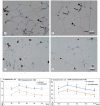
Photograph panel illustrating the appearance of the fat mass harvesting by the 2 lipofilling procedures (A), and the microanatomical appearance of the fat mass following the washing/fragmenting procedure (B) and the fat mass following the Coleman’s procedure (C). Note the wider presence of connective matrix and smaller size of adipocytes in the fat mass treated by the Coleman’s procedure in comparison to the washing/fragmenting procedure. Culture time: fourth week (T4), Line-graph illustrating morphometric analysis of connective tissue (D) and adipocytes size (E) performed along the different culture time. *= P< .05 vs. the other experimental group at the same time; §= P< .05 vs. the other experimental group at the same time and previous culture time of the same experimental group; a= P< .05 vs. previous culture time of the same experimental group.
Figure 2.
Figure 2.
Microphotograph panel illustrating the CD31 immunoreactive vessels profile occurring in the fat mass following the Coleman’s procedure (A) and the washing/fragmenting procedure (B). Culture time: third week (T3). Line-graph illustrating morphometric analysis of the CD31 immunoreactive vessels profile occurrence along the different culture time (C). Statistical significance symbols as in Figure 1.
Figure 3.
Figure 3.
Microphotograph panel illustrating the presence of CD31 immunoreactive leukocytes in the fat mass following the Coleman’s procedure (A), and in the washing/fragmenting procedure (B); CD68 immunoreactive macrophages in the fat mass following the Coleman’s procedure (C), and in the washing/fragmenting procedure (D). Culture time: first week (T1). Arrows, indicate the immunopositive vessels profile, arrowheads, indicate immunopositive leukocytes. Line-graph illustrating the analysis of the number of CD31 (E) and CD68 (F) immunoreactive leukocytes along the different culture time. Statistical significance symbols as in Figure 1.
Figure 4.
Figure 4.
Microphotograph panel illustrating the presence of CD8 immunoreactive leukocytes in the fat mass following the Coleman’s procedure (A), and in the washing/fragmenting procedure (B); Ki-67 immunoreactivity in the fat mass following the Coleman’s procedure (C), and in the washing/fragmenting procedure (D). Culture time: second week (T2). No differences have been observed between the 2 harvesting procedures.
Figure 5.
Figure 5.
Microphotograph panel illustrating M2-type macrophages (immunohistochemically detected) occurring in the fat mass following the Coleman’s procedure (A) and the washing/fragmenting procedure (B). Culture time: first week (T1). Line-graph illustrating the analysis along the different culture time of the number of M2-type macrophages (C). Statistical significance symbols as in Figure 1.
Figure 6.
Figure 6.
Microphotograph panel illustrating catalase detection and distribution in the fat mass following the Coleman’s procedure (A, B) and the washing/fragmenting procedure (C, D). Culture time: second week (T2). Line-graphs illustrating densitometric analysis of catalase reaction in adipocytes (E), immune cells (F), stroma environment (G), along the different culture time. Asterisk= adipocytes border; arrow= stromal environment; arrowhead= immune cells. Statistical significance symbols as in Figure 1.

References

    1. Billings E, Jr., May JW, Jr.. Historical review and present status of free fat graft auto transplantation in plastic and reconstructive surgery. Plast Reconstr Surg 1989;83:368-381. doi:10.1097/00006534-198902000-00033. - DOI - PubMed
    1. Bircoll M. Cosmetic breast augmentation utilizing autologous fat and liposuction technique. Plast Reconstr Surg. 1987;79:267-271. 10.1097/00006534-198702000-00022 - DOI - PubMed
    1. Debald M, Pech T, Kaiser C, Keyver-Paik M-D. Lipofilling effects after breast cancer surgery in post-radiation patients: an analysis of results and algorithm proposal. Eur J Plast Surg 2017;40:447-454. doi:10.1007/s00238-017-1311-1. - DOI - PMC - PubMed
    1. Gentile P, De Angelis B, Di Pietro V, et al. Gentle Is Better: The Original “Gentle Technique” for Fat Placement in Breast Lipofilling. J Cutan Aesthetic Surg. 2018;11:120-126. 10.4103/JCAS.JCAS_24_18 - DOI - PMC - PubMed
    1. Coleman S, Saboeiro A. Fat grafting to the breast revisited: safety and efficacy. Plast Reconstr Surg. 2007;119:775-785. 10.1097/01.prs.0000252001.59162.c9 - DOI - PubMed

Publication types