. 2021 Dec 11;26(1):52.
doi: 10.1186/s11658-021-00288-3.

MiR-26b-5p inhibits cell proliferation and EMT by targeting MYCBP in triple-negative breast cancer

Affiliations

MiR-26b-5p inhibits cell proliferation and EMT by targeting MYCBP in triple-negative breast cancer

Sugang Ma et al. Cell Mol Biol Lett. .

Abstract

Background: The study was designed to elucidate the association and functional roles of miR-26b-5p and c-MYC binding protein (MYCBP) in triple-negative breast cancer (TNBC).

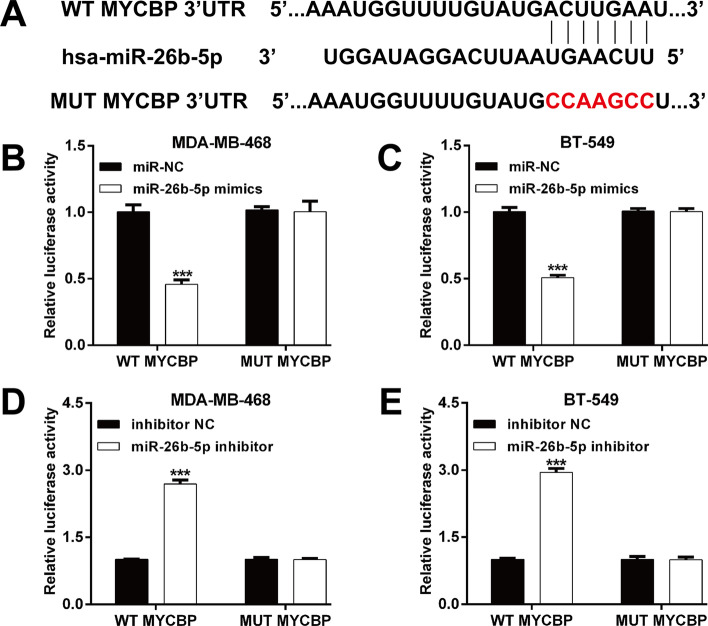
Method: Luciferase reporter assay was used to confirm the relationship between miR-26b-5p and MYCBP in TNBC cells. The expression levels of miR-26b-5p and MYCBP in tissue specimens and cell lines were determined using reverse transcription-quantitative PCR. Cell proliferation, migration and invasion were assessed using CCK-8 assay, colony formation and transwell assay.

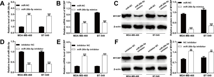
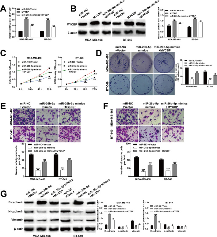
Results: We first observed that miR-26b-5p directly targets the 3'-UTR of MYCBP to inhibit MYCBP expression in MDA-MB-468 and BT-549 cells. The expression of miR-26b-5p was inversely correlated with MYCBP expression in TNBC tissues. We further demonstrated that MYCBP knockdown suppressed the proliferation, migration and invasion of TNBC cells. Furthermore, MYCBP overexpression counteracted the suppressive effect of miR-26b-5p on TNBC cell behaviors. Western blot analysis demonstrated that the E-cadherin protein level was increased, while protein levels of N-cadherin and vimentin were decreased in cells transfected with miR-26b-5p, which were all reversed by ectopic expression of MYCBP.

Conclusions: In summary, our findings revealed the tumor suppressive role of miR-26b-5p in regulating TNBC cell proliferation and mobility, possibly by targeting MYCBP.

Keywords: EMT; MYCBP; Triple-negative breast cancer; miR-26b-5p.


Conflict of interest statement

The authors declare that they have no competing interests.

Figures

Fig. 1
Fig. 1
MYCBP is a direct target of miR-26b-5p. A The binding site for miR-940 in the 3-′UTR of FOXO3 predicted by bioinformatics was presented. Luciferase reporter assay evaluation of the interaction between miR-26b-5p and the 3′-UTR of MYCBP mRNA. Luciferase activity was determined 48 h after co-transfection of luciferase constructs containing the wild-type (WT) or a mutated (MUT) 3′-UTR of MYCBP mRNA and miR-26b-5p mimics or miR-NC in MDA-MB-468 (B) and BT-549 (C) cells. Luciferase activity was determined 48 h after co-transfection of luciferase constructs containing the WT or a MUT 3′-UTR of MYCBP mRNA and miR-26b-5p inhibitor or inhibitor NC in MDA-MB-468 (D) and BT-549 (E) cells. Data represent the mean ± SD of three independent experiments. ***p < 0.001, compared with miR-NC or inhibitor NC
Fig. 2
Fig. 2
miR-26b-5p regulation of endogenous MYCBP expression. The expression levels of A miR-26b-5p, B MYCBP mRNA and C MYCBP protein were determined in MDA-MB-468 and BT-549 cells after transfection with miR-26b-5p mimics or miR-NC. The expression levels of D miR-26b-5p, E MYCBP mRNA and F MYCBP protein were determined in MDA-MB-468 and BT-549 cells after transfection with miR-26b-5p inhibitor or inhibitor NC. Data represent the mean ± SD of three independent experiments. ***p < 0.001, compared with miR-NC or inhibitor NC
Fig. 3
Fig. 3
Expression of miR-26b-5p and MYCBP in TNBC patients. A MiR-26b-5p levels were significantly lower in TNBC tissues (n = 60) than in adjacent nontumor tissues (ANT, n = 26). B The expression of miR-26b-5p in 20 pairs of TNBC tissues and matched ANT was determined using PCR assay. C MYCBP levels were significantly higher in TNBC tissues (n = 60) than in ANT (n = 26). D The expression of MYCBP in 20 pairs of TNBC tissues and matched ANT was determined using PCR assay. E The correlation between miR-26b-5p and MYCBP expression in TNBC tissues was determined by Spearman’s correlation analysis
Fig. 4
Fig. 4
Silencing of MYCBP inhibited TNBC cell proliferation, migration and invasion. Transfection efficiency of si-MYCBP#1 or si-MYCBP#2 in MDA-MB-468 and BT-549 cells was detected by PCR assay (A) and western blot analysis (B). C CCK-8 assay was used to determine the cell viability of MDA-MB-468 and BT-549 cells after transfection with si-MYCBP#1, si-MYCBP#2 or si-NC. D The influence of MYCBP knockdown on cell proliferation of MDA-MB-468 and BT-549 cells was detected by colony formation assay. MYCBP knockdown decreased the number of migrated (E) and invasive (F) cells. Magnification, ×200; scale bar, 100 μm; data are shown as mean ± SD (n = 3). *p < 0.05, **p < 0.01 and ***p < 0.001 compared with si-NC group
Fig. 5
Fig. 5
MYCBP overexpression counteracted the suppressive effect of miR-26b-5p on TNBC cell behaviors. MDA-MB-468 and BT-549 cells were co-transfected with MYCBP overexpressing plasmid and miR-26b-5p mimics. The expression levels of MYCBP mRNA (A) and protein (B) in MDA-MB-468 and BT-549 cells after indicated transfection were determined by PCR and western blot assays. C Cell viability was determined using CCK-8 assay in MDA-MB-468 and BT-549 cells after indicated transfection. D The number of colonies in MDA-MB-468 and BT-549 cells after indicated transfection. Cell migration (E) and invasion (F) were assessed by transwell assay in MDA-MB-468 and BT-549 cells after indicated transfection. Magnification, ×200; scale bar, 100 μm; data are shown as mean ± SD (n = 3). ***p < 0.001 compared with miR-NC + Vector group; #p < 0.05, ##p < 0.01, ###p < 0.001 compared with MYCBP or miR-26b-5p mimics; G The protein levels of E-cadherin, N-cadherin and vimentin were measured by western blot analysis in MDA-MB-468 and BT-549 cells after indicated transfection

References

    1. Zhai Q, Li H, Sun L, Yuan Y, Wang X. Identification of differentially expressed genes between triple and non-triple-negative breast cancer using bioinformatics analysis. Breast Cancer. 2019;26(6):784–791. doi: 10.1007/s12282-019-00988-x. - DOI - PubMed
    1. Bianchini G, Balko JM, Mayer IA, Sanders ME, Gianni L. Triple-negative breast cancer: challenges and opportunities of a heterogeneous disease. Nat Rev Clin Oncol. 2016;13(11):674–90. doi: 10.1038/nrclinonc.2016.66. - DOI - PMC - PubMed
    1. Guestini F, McNamara KM, Ishida T, Sasano H. Triple negative breast cancer chemosensitivity and chemoresistance: current advances in biomarkers indentification. Expert Opin Ther Targets. 2016;20(6):705–720. doi: 10.1517/14728222.2016.1125469. - DOI - PubMed
    1. Ambros V. The functions of animal microRNAs. Nature. 2004;431(7006):350–355. doi: 10.1038/nature02871. - DOI - PubMed
    1. Bartel DP. MicroRNAs: genomics, biogenesis, mechanism, and function. Cell. 2004;116(2):281–297. doi: 10.1016/S0092-8674(04)00045-5. - DOI - PubMed

MeSH terms