Review
. 2022 Mar;21(1):10-18.
doi: 10.1016/j.clcc.2021.10.007. Epub 2021 Nov 14.

MRI for Rectal Cancer: Staging, mrCRM, EMVI, Lymph Node Staging and Post-Treatment Response

Affiliations
Review

MRI for Rectal Cancer: Staging, mrCRM, EMVI, Lymph Node Staging and Post-Treatment Response

David D B Bates et al. Clin Colorectal Cancer. 2022 Mar.

Abstract

Rectal cancer is a relatively common malignancy in the United States. Magnetic resonance imaging (MRI) of rectal cancer has evolved tremendously in recent years, and has become a key component of baseline staging and treatment planning. In addition to assessing the primary tumor and locoregional lymph nodes, rectal MRI can be used to help with risk stratification by identifying high-risk features such as extramural vascular invasion and can assess treatment response for patients receiving neoadjuvant therapy. As the practice of rectal MRI continues to expand further into academic centers and private practices, standard MRI protocols, and reporting are critical. In addition, it is imperative that the radiologists reading these cases work closely with surgeons, medical oncologists, radiation oncologists, and pathologists to ensure we are providing the best possible care to patients. This review aims to provide a broad overview of the role of MRI for rectal cancer.

Keywords: Cancer staging; MRI; Radiology; Rectal cancer.


Conflict of interest statement

Disclosure The authors have stated that they have no conflicts of interest.

Figures
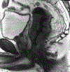
Figure 1:
Figure 1:
47 year-old male with newly diagnosed rectal adenocarcinoma for baseline staging. (a) Sagittal T2 weighted image of the mid to low rectal tumor (arrowheads), with an enlarged suspicious superior rectal lymph node (dashed arrow). (b) Axial oblique T2 weighted image through the lower part of the tumor shows extramural extension up to 0.8 cm (dashed line), conferring T3 disease. Enlarged, rounded heterogeneous lymph nodes in the mesorectum (c, arrowheads) and superior rectal chain (d, arrowheads) are consistent with metastatic total mesocolic excision (TME) nodes.
Figure 1:
Figure 1:
47 year-old male with newly diagnosed rectal adenocarcinoma for baseline staging. (a) Sagittal T2 weighted image of the mid to low rectal tumor (arrowheads), with an enlarged suspicious superior rectal lymph node (dashed arrow). (b) Axial oblique T2 weighted image through the lower part of the tumor shows extramural extension up to 0.8 cm (dashed line), conferring T3 disease. Enlarged, rounded heterogeneous lymph nodes in the mesorectum (c, arrowheads) and superior rectal chain (d, arrowheads) are consistent with metastatic total mesocolic excision (TME) nodes.
Figure 2:
Figure 2:
60 year-old male with newly diagnosed rectal adenocarcinoma for baseline staging. (a) Axial oblique T2-weighted image through the mid rectum shows a circumferential tumor (dashed arrow), with the mesorectal fascia noted (arrowheads). (b) More inferiorly in the rectum, there is extramural extension of tumor with multifocal involvement of the mesorectal fascia (arrowheads).
Figure 2:
Figure 2:
60 year-old male with newly diagnosed rectal adenocarcinoma for baseline staging. (a) Axial oblique T2-weighted image through the mid rectum shows a circumferential tumor (dashed arrow), with the mesorectal fascia noted (arrowheads). (b) More inferiorly in the rectum, there is extramural extension of tumor with multifocal involvement of the mesorectal fascia (arrowheads).
Figure 3:
Figure 3:
54 year-old male with rectal adenocarcinoma. (a) Axial oblique T2-weighted image through the rectum shows intermediate signal tumor expanding a mesorectal vein (arrowhead). The presence of tumor within the vein is better appreciated on coronal oblique T2-weighted images (b, c; arrowheads). This extramural vascular invasion (EMVI) extended to the right internal iliac vein (not shown).
Figure 3:
Figure 3:
54 year-old male with rectal adenocarcinoma. (a) Axial oblique T2-weighted image through the rectum shows intermediate signal tumor expanding a mesorectal vein (arrowhead). The presence of tumor within the vein is better appreciated on coronal oblique T2-weighted images (b, c; arrowheads). This extramural vascular invasion (EMVI) extended to the right internal iliac vein (not shown).
Figure 4:
Figure 4:
60 year-old male with rectal adenocarcinoma. (a) Coronal oblique T2 weighted image through the rectum shows a low rectal mass (dashed arrow) and a suspicious left obturator lymph node (arrowhead). (b) Axial oblique T2 image through the tumor (arrowhead) does not show definite extramural extension, but the 0.6 cm left obturator node (c, arrowhead) demonstrates a rounded morphology, heterogeneous signal and an irregular border, suspicious for nodal metastasis despite its small size.
Figure 4:
Figure 4:
60 year-old male with rectal adenocarcinoma. (a) Coronal oblique T2 weighted image through the rectum shows a low rectal mass (dashed arrow) and a suspicious left obturator lymph node (arrowhead). (b) Axial oblique T2 image through the tumor (arrowhead) does not show definite extramural extension, but the 0.6 cm left obturator node (c, arrowhead) demonstrates a rounded morphology, heterogeneous signal and an irregular border, suspicious for nodal metastasis despite its small size.
Figure 5:
Figure 5:
61 year-old male with rectal adenocarcinoma status post total neoadjuvant therapy with complete clinical response. (a) Axial oblique T2 weighted image shows semi-circumferential low signal scar in the anterior rectum with adjacent desmoplastic spiculations in the mesorectal fat. (b) A follow-up MRI 9 months later shows a new small intermediate signal mass in the scar (arrowhead), with high signal on b1500 s/mm2 diffusion weighted imaging (c, arrowhead) and corresponding hypointense signal on the apparent diffusion coefficient map (d, arrowhead), consistent with tumor re-growth in the scar.
Figure 5:
Figure 5:
61 year-old male with rectal adenocarcinoma status post total neoadjuvant therapy with complete clinical response. (a) Axial oblique T2 weighted image shows semi-circumferential low signal scar in the anterior rectum with adjacent desmoplastic spiculations in the mesorectal fat. (b) A follow-up MRI 9 months later shows a new small intermediate signal mass in the scar (arrowhead), with high signal on b1500 s/mm2 diffusion weighted imaging (c, arrowhead) and corresponding hypointense signal on the apparent diffusion coefficient map (d, arrowhead), consistent with tumor re-growth in the scar.

References

    1. Al-Sukhni E, Milot L, Fruitman M, Beyene J, Victor JC, Schmocker S, Brown G, McLeod R, Kennedy E. Diagnostic accuracy of MRI for assessment of T category, lymph node metastases, and circumferential resection margin involvement in patients with rectal cancer: a systematic review and meta-analysis. Ann Surg Oncol. 2012;19(7):2212–23. - PubMed
    1. Costa-Silva L, Brown G. Magnetic resonance imaging of rectal cancer. Magn Reson Imaging Clin N Am. 2013;21(2):385–408. - PubMed
    1. Furey E, Jhaveri KS. Magnetic resonance imaging in rectal cancer. Magn Reson Imaging Clin N Am. 2014;22(2):165–90, v-vi. - PubMed
    1. Beets-Tan RGH, Lambregts DMJ, Maas M, Bipat S, Barbaro B, Curvo-Semedo L, Fenlon HM, Gollub MJ, Gourtsoyianni S, Halligan S, Hoeffel C, Kim SH, Laghi A, Maier A, Rafaelsen SR, Stoker J, Taylor SA, Torkzad MR, Blomqvist L. Magnetic resonance imaging for clinical management of rectal cancer: Updated recommendations from the 2016 European Society of Gastrointestinal and Abdominal Radiology (ESGAR) consensus meeting. Eur Radiol. 2018;28(4):1465–75. - PMC - PubMed
    1. Taylor FG, Swift RI, Blomqvist L, Brown G. A systematic approach to the interpretation of preoperative staging MRI for rectal cancer. AJR Am J Roentgenol. 2008;191(6):1827–35. - PubMed

Publication types

MeSH terms