. 2022 Jan:175:106030.
doi: 10.1016/j.phrs.2021.106030. Epub 2021 Dec 9.

Toll-like receptor 4 mutation protects the kidney from Ang-II-induced hypertensive injury

Affiliations

Toll-like receptor 4 mutation protects the kidney from Ang-II-induced hypertensive injury

Suravi Majumder et al. Pharmacol Res. 2022 Jan.

Abstract

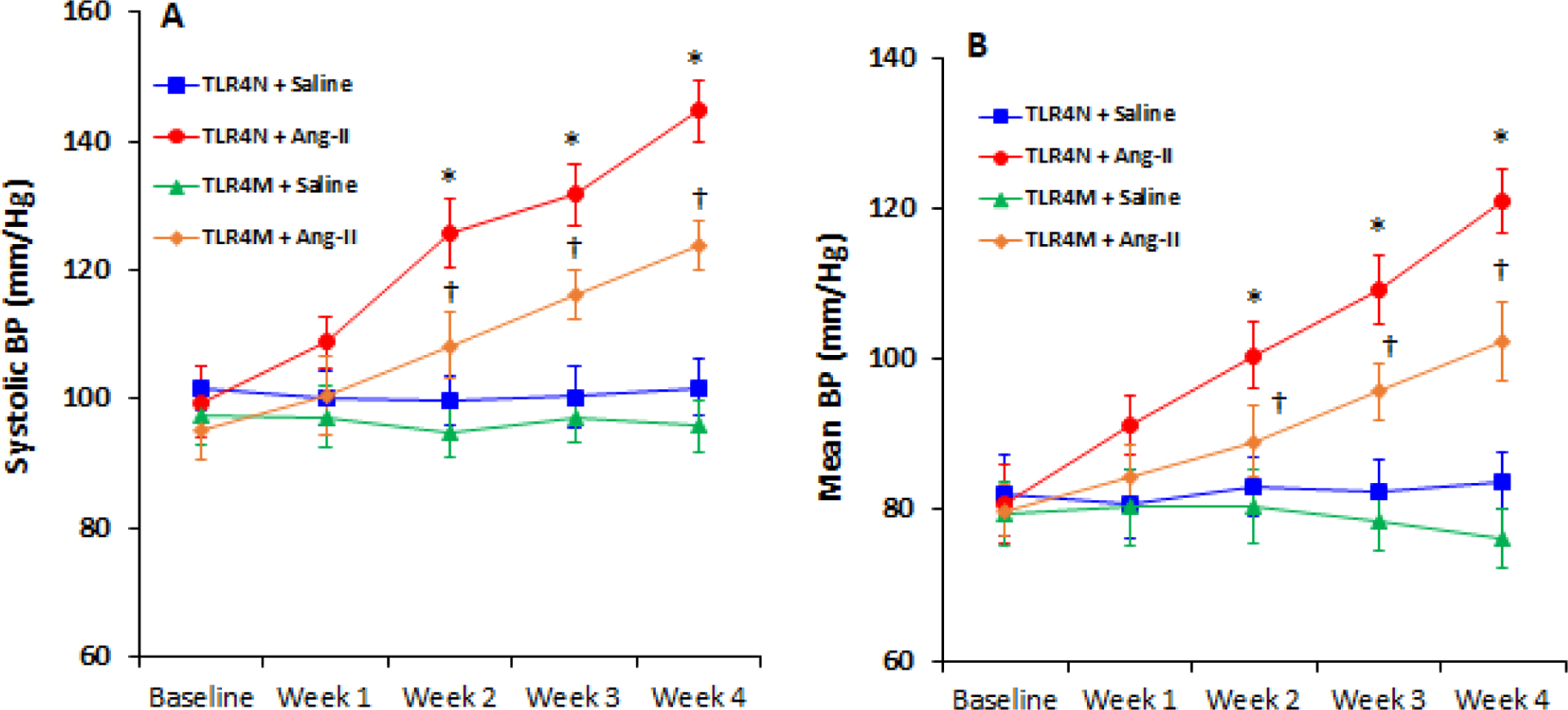
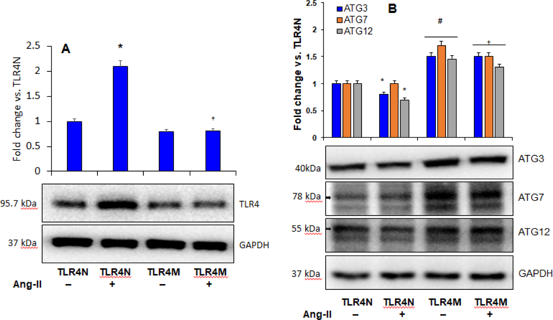
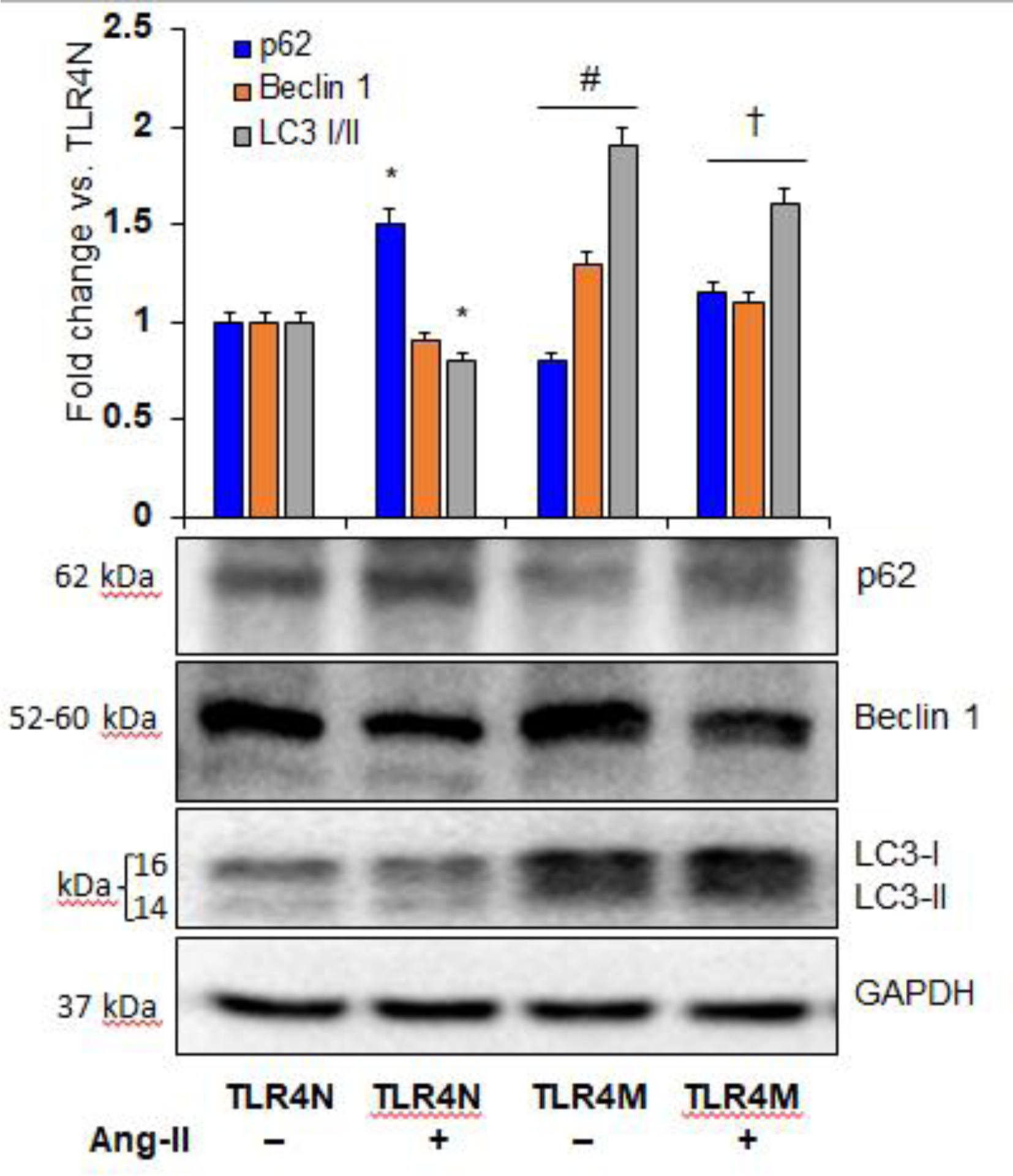
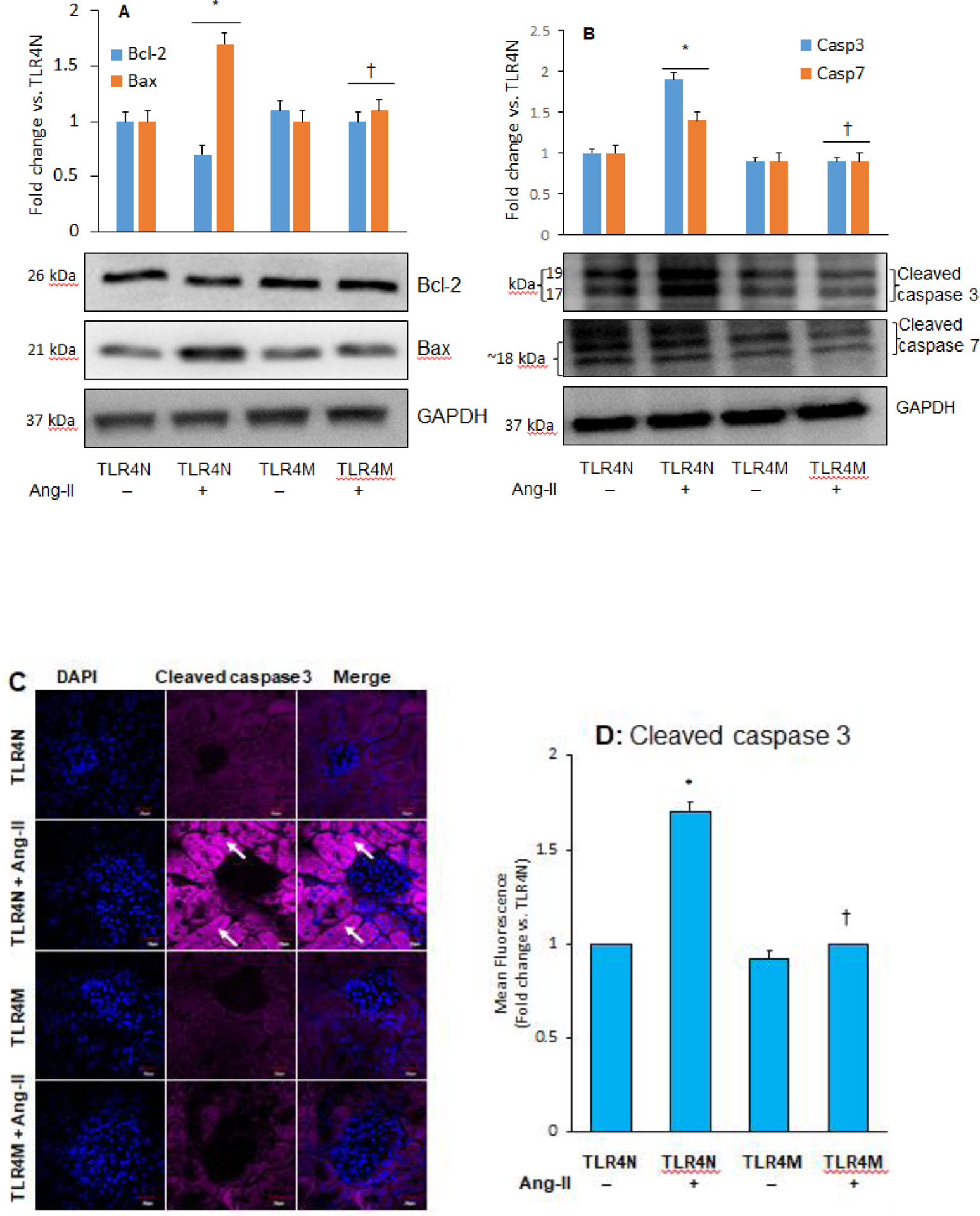
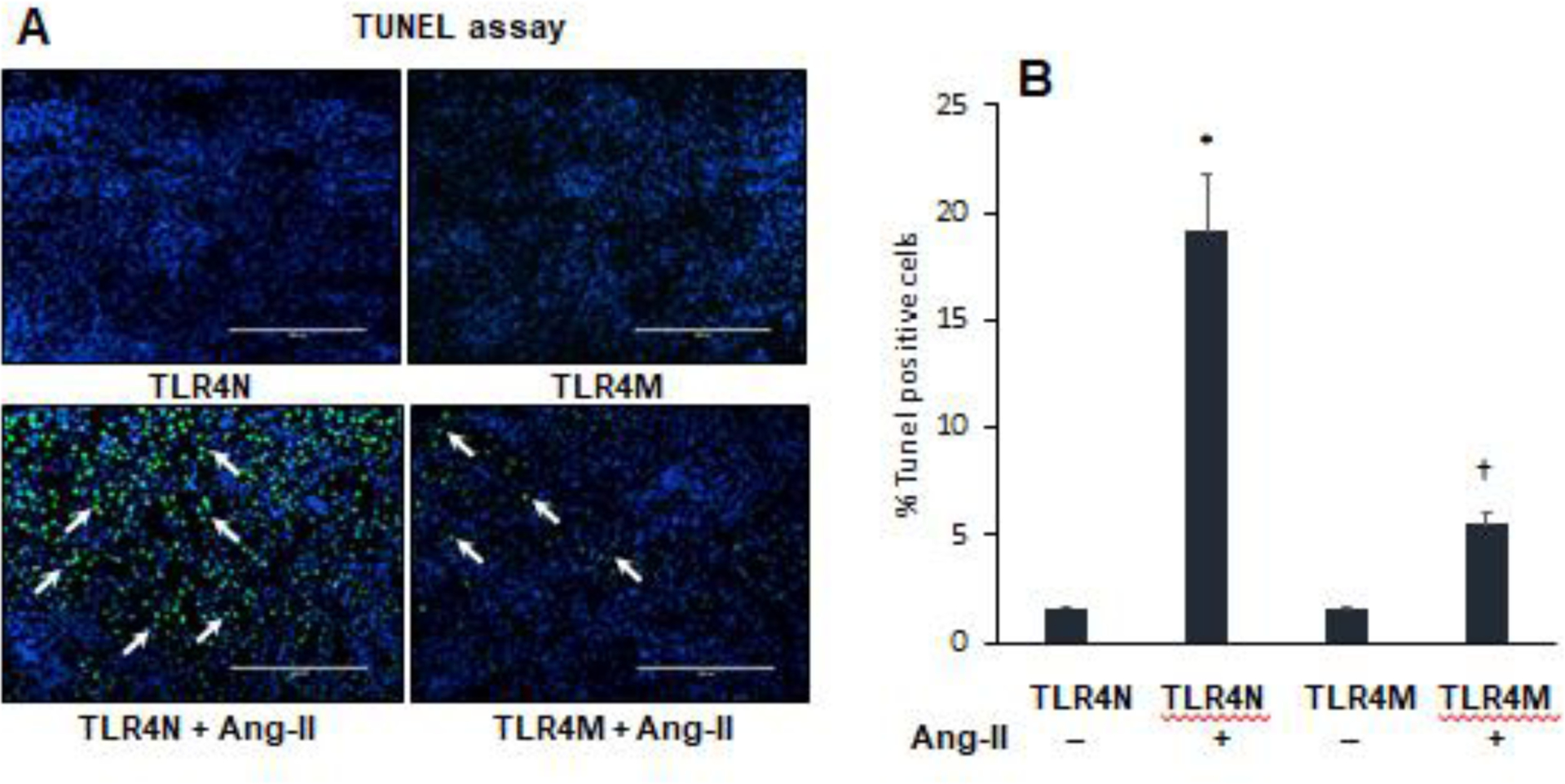
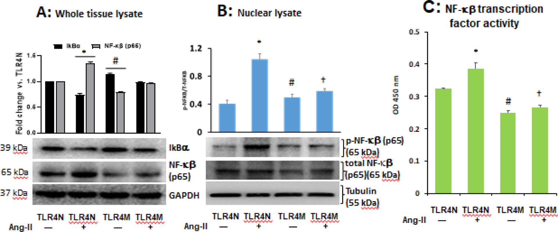
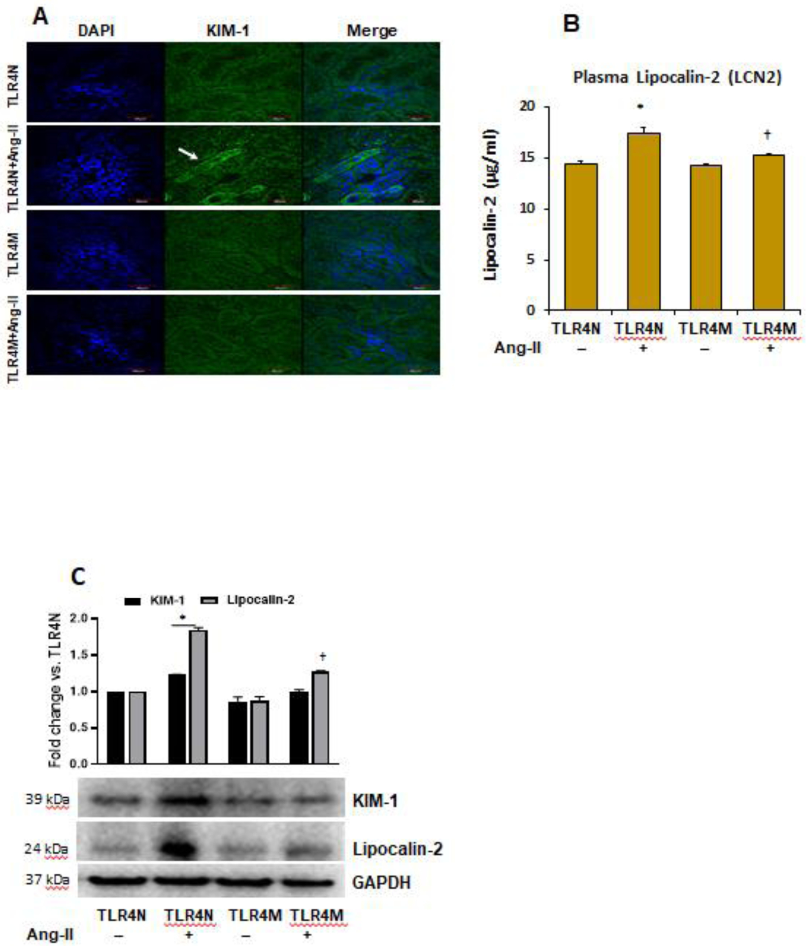
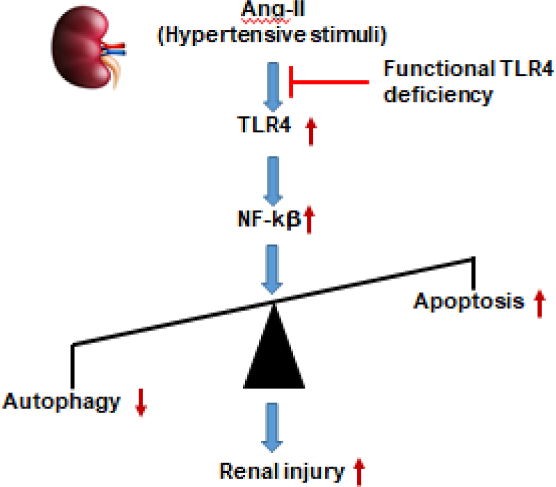
Cellular autophagy is a protective mechanism where cells degrade damaged organelles to maintain intracellular homeostasis. Apoptosis, on the other hand, is considered as programmed cell death. Interestingly, autophagy inhibits apoptosis by degrading apoptosis regulators. In hypertension, an imbalance of autophagy and apoptosis regulators can lead to renal injury and dysfunction. Previously, we have reported that toll-like receptor 4 (TLR4) mutant mice are protective against renal damage, in part, due to reduced oxidative stress and inflammation. However, the detailed mechanism remained elusive. In this study, we tested the hypothesis of whether TLR4 mutation reduces Ang-II-induced renal injury by inciting autophagy and suppressing apoptosis in the hypertensive kidney. Male mice with normal TLR4 expression (TLR4N, C3H/HeOuJ) and mutant TLR4 (TLR4M, C3H/HeJLps-d) aged 10-12 weeks were infused with Ang-II (1000 ng/kg/d) for 4 weeks to create hypertension. Saline infused appropriate control were used. Blood pressure was increased along with increased TLR4 expression in TLR4N mice receiving Ang-II compared to TLR4N control. Autophagy was downregulated, and apoptosis was upregulated in TLR4N mice treated with Ang-II. Also, kidney injury markers plasma lipocalin-2 (LCN2) and kidney injury molecule 1 (KIM-1) were upregulated in TLR4N mice treated with Ang-II. Besides, increased nuclear translocation and activity of NF-kB were measured in Ang-II-treated TLR4N mice. TLR4M mice remained protected against all these insults in hypertension. Together, these results suggest that Ang-II-induced TLR4 activation suppresses autophagy, induces apoptosis and kidney injury through in part by activating NF-kB signaling, and TLR4 mutation protects the kidney from Ang-II-induced hypertensive injury.

Keywords: Apoptosis; Autophagy; Hypertension; Injury; Kidney; TLR4.


Conflict of interest statement

Disclosure of Potential Conflicts of Interest

The authors declare no conflicts of interest, financial or otherwise.

Figures

Fig. 1.
Fig. 1.
TLR4 mutant mice exhibited resistance to Ang-II induced hypertension. Time course blood pressure (BP) was measured following Ang-II and saline infusion through osmotic mini-pump. (A) systolic and (B) mean BP in TLR4N and TLR4M mice with or without Ang-II. Data presented as mean ± SEM, n = 5/saline (control) groups, and n=6/Ang-II treated groups: *P < 0.05 vs. TLR4N+saline (control), and †P < 0.05 vs. TLR4N+Ang-II.
Fig. 2.
Fig. 2.
A) Ang-II increased TLR4 expression in TLR4N mice. Representative Western blot image of TLR4 protein expression in the kidney. Bar graph shows fold change of protein expression. Data presented as mean ± SEM, n=5/saline (control) groups, and n=6/Ang-II treated groups: *P < 0.05 vs. TLR4N+saline (control), and †P<0.05 vs. TLR4N+Ang-II. B) TLR4 mutation protected Ang-II-mediated inhibition of autophagy in the kidney. Representative Western blot images of autophagy marker proteins in the kidney. Bar graph represents fold change of protein expression. Data presented as mean ± SEM, n=5/saline (control) groups, and n=6/Ang-II treated groups: * and #P< 0.05 vs. TLR4N+saline (control), and †P< 0.05 vs. TLR4N+Ang-II of respective ATGs.
Fig. 3.
Fig. 3.
Ang II-mediated autophagy inhibition in TLR4N kidney was restored in TLR4M kidney. Representative Western blot images of autophagy marker proteins p62, Beclin 1, and LC3- I/II expression in the kidney. Respective bar graphs represent fold change of protein expression. Data presented as mean ± SEM, n=5/saline (control) groups, and n=6/Ang-II treated groups: * and #P< 0.05 vs. TLR4N+saline (control), and †P<0.05 vs. TLR4N+Ang-II of respective proteins.
Fig. 4.
Fig. 4.
TLR4 mutation decreased Ang-II-induced apoptosis in the kidney. Representative Western blot images for apoptosis marker proteins (A) Bcl-2 and Bax, and (B) cleaved caspase 3 and 7 in the kidney. Bar graphs represents fold change of protein expression, Data presented as mean ± SEM, n=5/saline (control) groups, and n=6/Ang-II treated groups: *P<0.05 vs. TLR4N+saline (control), and †P< 0.05 vs. TLR4N+Ang-II of respective proteins. C) Increased cleaved caspase 3 expression in hypertensive kidney was normalized in TLR4 mutant mice. Arrows indicate cleaved caspase 3 expression in renal tubules. Representative images from random fields (5 fields/slide/animal), Magnification 60X. Scale bar 20 μM. (D) Bar graph represents fold change of cleaved caspase 3 expression in the immunostained sections. Data presented as mean ± SEM, n=5/saline (control) groups, and 6/Ang-II treated groups: *P<0.05 vs. TLR4N+saline (control), and †P<0.05 vs. TLR4N+Ang-II.
Fig. 5.
Fig. 5.
TLR4 mutant mice were protected from apoptotic cells death in hypertensive kidney. (A) Bright green cells are TUNEL positive cells (arrow heads) in the kidney section. Representative images from random fields (5 fields/slide/animal). Magnification 20X. Scale bar 200 μM. (B) Bar graph indicates percent change of number of dead cells. Data presented as mean ± SEM, n=5/saline (control) groups, and n=6/Ang-II treated groups: *P<0.05 vs. TLR4N+saline (control), and †P<0.05 vs. TLR4N+Ang-II.
Fig. 6.
Fig. 6.
Ang-II-induced diminished IkBα and upregulated NF-κB (p65) expression in the TLR4N kidney was normalized in TLR4M kidney. (A) Representatives immunoblot images of IkBα and NF-κB (p65). GAPDH used as loading control. (B) TLR4 mutation prevented nuclear translocation of Ang-II-induced p-NF-κB (p65) in the kidney tissue. Tubulin was used as loading control; p-NF-κB p65, phospho-NF-κB (p65). C) TLR4 mutation prevented Ang-II-induced NF-κB transcription factor activity in kidney tissue. All bar graphs represent fold change compared to TLR4N+saline (control). Data presented as mean ± SEM, n=5/saline (control) groups, and n=6/Ang-II treated groups: * and #P<0.05 vs. TLR4N+saline (control), and P<0.05 vs. TLR4N+Ang-II.
Fig. 7.
Fig. 7.
TLR4 mutation protected kidney from injury in Ang-II hypertension. (A) Kidney injury molecule-1 (KIM-1) was detected by immunostaining. Representative images from random fields (5 fields/slide/animal). Magnification 60X. Scale bar 40 μM. (B) plasma Lipocalin-2 (LCN2) was measured by ELISA. Data represent mean ± SEM, n=5/saline (control) groups, and n=6/Ang-II treated groups: *P<0.05 vs. TLR4N+saline (control), and †P<0.05 vs. TLR4N+Ang-II. (C) Western blot images of kidney tissue KIM-1 and Lipocalin-2 expression. GAPDH was used as loading control. Bar graphs represent fold change of protein expression. Data presented as mean ± SEM, n=5/saline (control) groups, and n=6/Ang-II treated groups: *P<0.05 vs. TLR4N+saline (control), and †P< 0.05 vs. TLR4N+Ang-II of respective proteins.
Fig. 8.
Fig. 8.
Schematic of findings. Ang-II-induced activation of TLR4 upregulates NF-κB in the kidney leading to impaired homeostasis of autophagy and apoptosis resulting in renal injury. Mutation of TLR4 protects Ang-II-induced renal injury by inducing autophagy and inhibiting apoptosis in the kidney during hypertension.

References

    1. Hill NR, Fatoba ST, Oke JL, Hirst JA, O’Callaghan CA, Lasserson DS, Hobbs FD, Global Prevalence of Chronic Kidney Disease - A Systematic Review and Meta-Analysis, PLoS One 11(7) (2016) e0158765. - PMC - PubMed
    1. Rai A, Narisawa M, Li P, Piao L, Li Y, Yang G, Cheng XW, Adaptive immune disorders in hypertension and heart failure: focusing on T-cell subset activation and clinical implications, J Hypertens 38(10) (2020) 1878–1889. - PubMed
    1. Biancardi VC, Bomfim GF, Reis WL, Al-Gassimi S, Nunes KP, The interplay between Angiotensin II, TLR4 and hypertension, Pharmacol Res 120 (2017) 88–96. - PubMed
    1. Pushpakumar S, Ren L, Kundu S, Gamon A, Tyagi SC, Sen U, Toll-like Receptor 4 Deficiency Reduces Oxidative Stress and Macrophage Mediated Inflammation in Hypertensive Kidney, Sci Rep 7(1) (2017) 6349. - PMC - PubMed
    1. Bondeva T, Roger T, Wolf G, Differential regulation of Toll-like receptor 4 gene expression in renal cells by angiotensin II: dependency on AP1 and PU.1 transcriptional sites, Am J Nephrol 27(3) (2007) 308–14. - PubMed

Publication types

MeSH terms