Review
. 2021 Dec 13;6(1):412.
doi: 10.1038/s41392-021-00823-w.

T cell receptor (TCR) signaling in health and disease

Affiliations
Review

T cell receptor (TCR) signaling in health and disease

Kinjal Shah et al. Signal Transduct Target Ther. .

Abstract

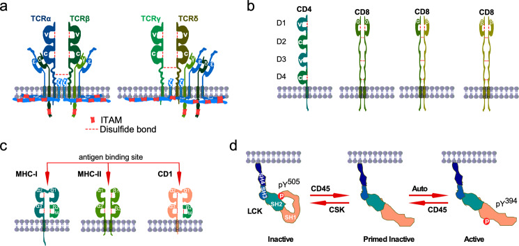
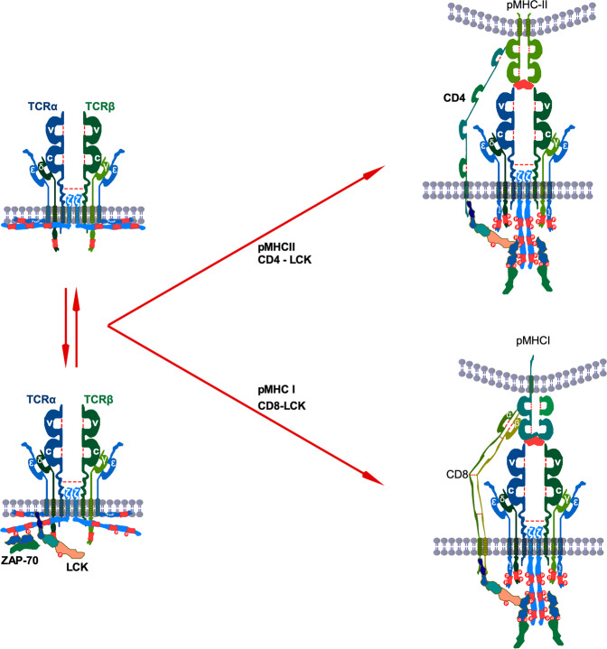
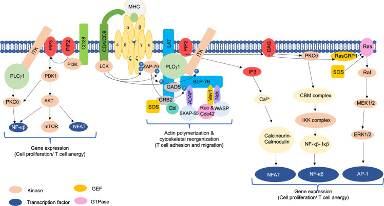
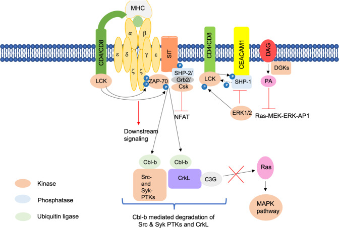
Interaction of the T cell receptor (TCR) with an MHC-antigenic peptide complex results in changes at the molecular and cellular levels in T cells. The outside environmental cues are translated into various signal transduction pathways within the cell, which mediate the activation of various genes with the help of specific transcription factors. These signaling networks propagate with the help of various effector enzymes, such as kinases, phosphatases, and phospholipases. Integration of these disparate signal transduction pathways is done with the help of adaptor proteins that are non-enzymatic in function and that serve as a scaffold for various protein-protein interactions. This process aids in connecting the proximal to distal signaling pathways, thereby contributing to the full activation of T cells. This review provides a comprehensive snapshot of the various molecules involved in regulating T cell receptor signaling, covering both enzymes and adaptors, and will discuss their role in human disease.


Conflict of interest statement

The authors declare no competing interests.

Figures

Fig. 1
Fig. 1. TCR components.
a TCRα/TCRβ and TCRγ/TCRδ heterodimers form complexes with the CD3 molecules. Heterodimers of CD3ε/CD3δ and CD3γ/CD3ε, and a homodimer of CD3ζ/CD3ζ form complexes with TCR dimers. TCR heterodimers contain intramolecular and intermolecular disulfide bonds. CD3 chains contain 10 ITAMs distributed in different CD3 molecules. The variable region (V) of TCR heterodimers recognize the antigen peptide-loaded on MHC (pMHC). In the absence of pMHC, the intracellular part of the CD3 molecules forms a close conformation in which ITAMs are inaccessible to the kinases for phosphorylation. b Coreceptor CD4 acts as a single molecule while CD8α and CD8β can form homodimers or heterodimers. c MCH-I consists of an α-chain containing three immunoglobulin domains (α1, α2, α3) and β2-microglobulin (β2m). MCH-2 is the heterodimer of an α chain and a β-chain containing two immunoglobulin domains (α1, α2, and β1, β2) in each chain. d LCK-loaded CD4 molecules bind to the MHC-II bound TCR (TCRα/TCRβ) complex. This allows LCK to phosphorylate two distinct sites on ITAMs. Then ZAP-70 interacts with the phosphotyrosine sites and mediates more tyrosine phosphorylation. CD4 and MHC-II interaction is mediated through the membrane-proximal α2 and β2 domains of MHC-II and the membrane-distal D1 domain of CD4.
Fig. 2
Fig. 2. TCR activation.
In resting T cells, CD3ζ and CD3ε remain membrane-embedded. Perhaps membrane-bound CD3ζ might be released to the cytosol, where free LCK induces tyrosine phosphorylation on at least two sites in ITAMs. This basal tyrosine phosphorylation creates docking sites for ZAP-70 interaction. After antigen engagement, the TCR complex recruits coreceptor-bound LCK that phosphorylates ZAP-70 and interacts with it through the SH2 domain facilitating tyrosine phosphorylation on other residues on ITAMs.
Fig. 3
Fig. 3. Positive regulation of T cell signaling.
The figure depicts the activation of various enzymes and adaptor molecules upon engagement of TCR with the MHC antigenic peptide complex. The phosphorylation events carried out are depicted as small, blue-colored circles. Black lines with arrows indicate activation.
Fig. 4
Fig. 4. Negative regulation of T cell signaling.
The figure depicts various adaptors and enzymes, like kinases and phosphatases, involved in negatively regulating TCR signaling. The phosphorylation events carried out are depicted as small, blue-colored circles. Black lines with arrows indicate activation. Dotted black lines with arrows indicate dephosphorylation events.
Fig. 5
Fig. 5. Schematic illustration of TCR-based immunotherapy.
T cells are isolated from the patient’s cancer tissue or peripheral blood and genetically modified by retroviral transduction to express antigen-specific TCR or CAR on T cells. Cells are then expanded ex vivo until sufficient cell numbers are achieved and reinfused into the patient’s body, where they can fight cancer cells.
Fig. 6
Fig. 6. Schematic representation of the tumor microenvironment (TME).
The immunosuppressive microenvironment induced by cancer-associated stromal cells modulates cancer progression and therapy resistance. Infiltration of immune cells, such as T reg cells, N2 neutrophils, tumor-associated macrophages, MDSC cells, the transformation of malignant fibroblasts, release of pro-inflammatory cytokines and chemokines, dysregulated vasculature and extracellular matrix remodeling, overexpression of negative immune-checkpoint regulators, metabolic status of the tumor including O2 and nutrients deprivation, the genetic composition of the tumor cells, all this heterogeneous ecosystem of the TME contributes to the tumor therapy resistance.

References

    1. Gorentla BK, Zhong XP. T cell receptor signal transduction in T lymphocytes. J. Clin. Cell Immunol. 2012;2012:5. - PMC - PubMed
    1. Marshall JS, Warrington R, Watson W, Kim HL. An introduction to immunology and immunopathology. Allergy Asthma Clin. Immunol. 2018;14:49. - PMC - PubMed
    1. Quang CT, Zaniboni B, Ghysdael J. A TCR-switchable cell death pathway in T-ALL. Oncoscience. 2017;4:17–18. - PMC - PubMed
    1. Zuniga-Pflucker JC. T-cell development made simple. Nat. Rev. Immunol. 2004;4:67–72. - PubMed
    1. Sallusto F, Geginat J, Lanzavecchia A. Central memory and effector memory T cell subsets: function, generation, and maintenance. Annu. Rev. Immunol. 2004;22:745–763. - PubMed

Publication types

MeSH terms

Substances