. 2022 Jan 15;11(1):bio059128.
doi: 10.1242/bio.059128. Epub 2022 Jan 18.

Redundant functions of the SLC5A transporters Rumpel, Bumpel, and Kumpel in ensheathing glial cells

Affiliations

Redundant functions of the SLC5A transporters Rumpel, Bumpel, and Kumpel in ensheathing glial cells

Kerem Yildirim et al. Biol Open. .

Abstract

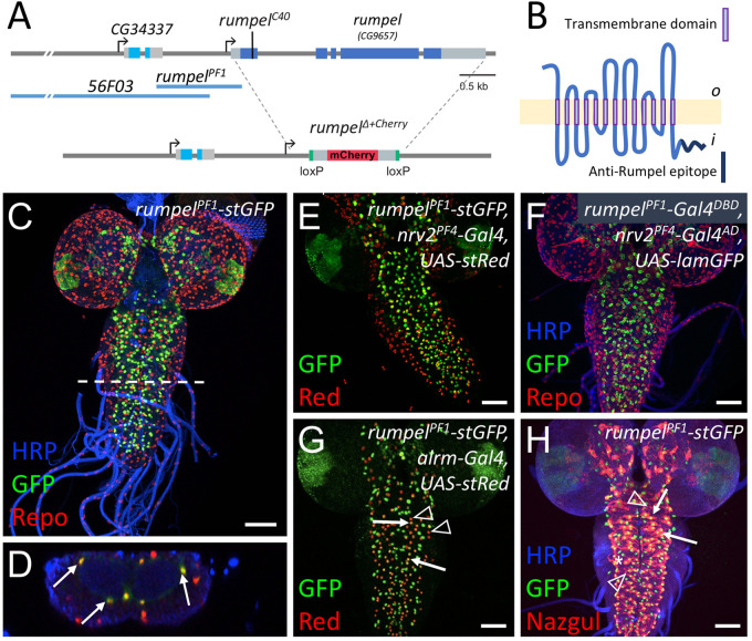
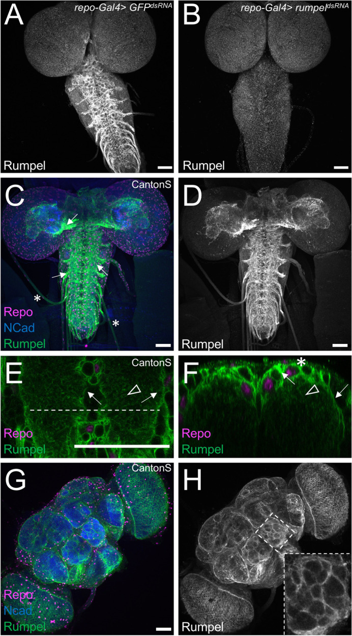
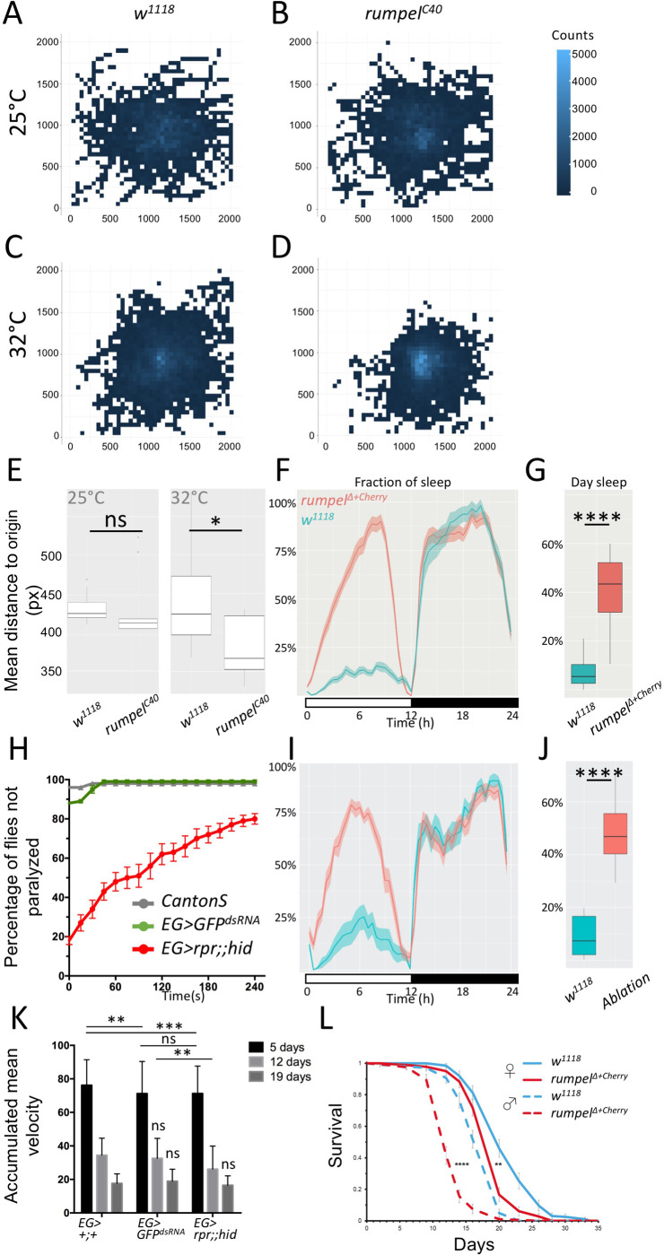
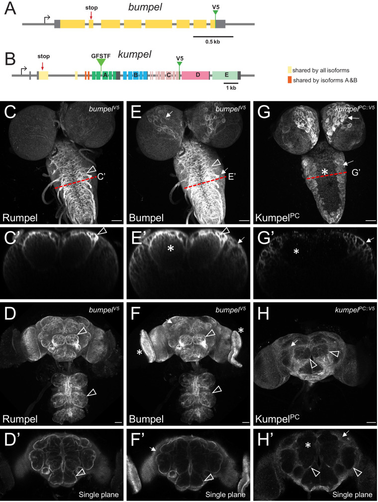
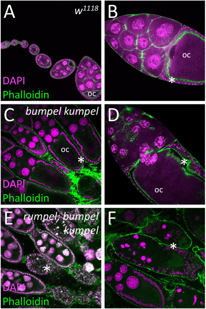
Neuronal processing is energy demanding and relies on sugar metabolism. To nurture the Drosophila nervous system, the blood-brain barrier forming glial cells take up trehalose from the hemolymph and then distribute the metabolic products further to all neurons. This function is provided by glucose and lactate transporters of the solute carrier (SLC) 5A family. Here we identified three SLC5A genes that are specifically expressed in overlapping sets of CNS glial cells, rumpel, bumpel and kumpel. We generated mutants in all genes and all mutants are viable and fertile, lacking discernible phenotypes. Loss of rumpel causes subtle locomotor phenotypes and flies display increased daytime sleep. In addition, in bumpel kumpel double mutants, and to an even greater extent in rumpel bumpel kumpel triple mutants, oogenesis is disrupted at the onset of the vitollegenic phase. This indicates a partially redundant function between these genes. Rescue experiments exploring this effect indicate that oogenesis can be affected by CNS glial cells. Moreover, expression of heterologous mammalian SLC5A transporters, with known transport properties, suggest that Bumpel and/or Kumpel transport glucose or lactate. Overall, our results imply a redundancy in SLC5A nutrient sensing functions in Drosophila glial cells, affecting ovarian development and behavior.

Keywords: CG42235; CG6723; CG9657; Drosophila; Ensheathing glia; Redundancy; SLC5A transporters.


Conflict of interest statement

Competing interests The authors declare no competing or financial interests.

Figures

Fig. 1.
Fig. 1.
The expression of SLC5 family members in the adult brain. (A–N) Single cell RNA sequencing data in the SCENIC representations of the 57 K scRNA seq data set (Davie et al., 2018). SCope analysis for the genes indicated in each bottom right corner is shown. Each dot represents a single cell. The color coding indicates the expression level. Red: strong expression, black: low expression. Grey: no expression. (A,B) repo expression marks glial cell clusters that can be assigned as perineurial (yellow), subperineurial (blue), cortex (grey), ensheathing (green) or astrocyte-like glial cells (orange) according to marker gene expression as shown in (C–H). (I–N) Expression of SLC5 family members that show expression in Drosophila glia. (O) Dendrogram of the evolutionary relationships of the different SLC5 family members of Drosophila. The color shading indicates expression in the respective glial cell type (see B). The scale bar represents 2×10⁵ years of evolutionary distance.
Fig. 2.
Fig. 2.
rumpel-PF1 induces an expression in the neuropil-associated glial cells. (A) Schematic representation of the rumpel (CG9657) locus on the X-chromosome. Exons are shown in boxes, rumpel coding exons are in dark blue, 56F03 and rumpel PF1 denote enhancer elements that direct expression in ensheathing glia. The position of the CRISPR-induced premature stop codon in amorphic allele (rumpelC40) and the rumpel locus replacement with attP-loxP-Cherry-loxP in (rumpelΔ+Cherry) is indicated. (B) The Rumpel protein is predicted to have 13 membrane (light yellow) spanning domains. The peptide sequence used to immunize rabbits is highlighted in dark blue. o, outside; i, inside. (C–E,G,H) Specimens are stained for promoter fragment induced expression of StingerGFP (stGFP, green). (E,G) RedStinger (stRed, red). (F) LaminGFP (lamGFP, green). (C,D) Glial nuclei are stained for Repo protein localization (red). (H) Astrocyte-like glial cells are stained for Nazgul protein localization (red). Neuronal membranes are shown in blue (HRP staining). (C) rumpel promoter fragment PF1 (rumpelPF1) induces stGFP expression in Repo positive cells in the third instar larval brain. White dashed line indicates the position of the orthogonal section shown in D. (D) Glial cells in the position of ensheathing glia are indicated by arrows. No expression is observed in surface associated glial cells. (E) rumpelPF1 induced stGFP expression overlaps with the nrv2 induced RedStinger expression. (F) Split Gal4 directed expression of LamGFP is found in ensheathing glial cells [rumpelPF1-Gal4DBD, nrv2PF4-Gal4AD, UAS-lamGFP]. (G) rumpelPF1 induced stGFP expression is found in some astrocyte-like glial cells labelled by alrm induced stRed expression (compare arrows with arrowheads). (H) rumpelPF1 induced stGFP expression in Nazgul positive astrocyte-like glial cells (arrows). The asterisk denotes ensheathing glial nuclei, the arrowhead denotes astrocytes not activating the rumpelPF1 enhancer. Scale bars: 50 µm.
Fig. 3.
Fig. 3.
Rumpel protein is expressed in the neuropil-associated glial cells. All specimens are stained for Repo localization to define glial nuclei (magenta), for N-Cadherin localization to visualize axonal and dendritic cell membranes (blue) and for Rumpel protein localization (green/grey). (A–F) Third instar larval brains and (G,H) adult brain. (A) In control animals [repo-Gal4, UAS-GFPdsRNA] Rumpel protein localizes around the neuropil. (B) Upon expression of rumpeldsRNA in the all-glial cells [repo-Gal4, UAS- rumpeldsRNAv43922] no Rumpel protein can be detected, demonstrating the specificity of the anti-Rumpel antibody. (C,D) Rumpel localization is observed surrounding the neuropil (arrows) in a position of the ensheathing glial cells. Very little Rumpel protein is found along larval nerves (asterisks). (E) Image of a single confocal plane through a third instar larval ventral nerve cord. Rumpel localizes to ensheathing glial cell membrane (arrow) and to cell processes of astrocyte-like glial cells (arrowhead). The dashed line indicates the position of the orthogonal section shown in F. (F) Rumpel localizes to ensheathing glial cells (arrows) and astrocytic processes in the neuropil (arrowhead). Note, the pronounced cortex-glial cell like ramifications of the ensheathing glia dorsally to the neuropil (asterisk). (G,H) Rumpel localizes around the neuropil in adult brains at a position of the ensheathing glia (inset: antennal lobe). Scale bars: 50 µm.
Fig. 4.
Fig. 4.
Behavioral analysis of rumpel. (A–E) 150 third instar larvae of the respective genotypes were recorded in groups of 15 animals for 3 min at 25°C or 32°C, as indicated. Larvae were always placed at the middle of the tracking plate. (A–D) For heatmap analyses, the 2048×2048 px image of the agar plate is divided in 50×50 px squares. The number of larval appearances per square is determined and indicated in blue shading using R. Darker blue colors indicate less frequent appearance, while lighter blue ones more. (A) Heatmap analysis of control w1118 larvae at 25°C. Wild-type larvae crawl in every direction and spread evenly on the agar plate at 25°C (indicated by fewer lighter blue squares). (B) Heatmap analysis of rumpelC40 larvae. rumpelC40 larvae shows wild type-like distribution on the agar plate at 25°C (indicated by similar number of lighter blue squares). (C) Wild-type larvae spread evenly on the agar plate at 32°C. (D) At 32°C rumpelC40 larvae spread less on the agar plate (indicated by more light blue squares in the middle). (E) Quantification of the mean distance to origin of wild-type versus rumpelC40 larvae at 25°C and 32°C. At 25°C no significant difference is indicated (Mann–Whitney U-test P>0.05, n=150). Mean distance to origins of wild type and rumpelC40 are 439.4 and 381.4 px, respectively at 32°C. Wild-type larvae spread significantly more on the agar plate at 32°C compared to rumpelC40 larvae (Mann–Whitney U-test P=0.023, n=150). (F) To monitor the effects of rumpel on sleep behavior rumpelΔ+cherry flies were backcrossed 10 times to white1118. The activity of 40 flies was tracked over 7 days in the ethoscope (Geissmann et al., 2017). (G) rumpelΔ+cherry flies sleep significantly more during the day (P=2e-16, Wilcoxon rank sum), whereas night sleep is not affected. (H) Heat shock assay of 5-day-old male and mated female flies of wild type [Canton S], [GMR83E12-Gal4AD; repo-Gal4DBD, UAS-GFPdsRNA], [UAS-rpr; GMR83E12-Gal4AD; repo-Gal4DBD, UAS-hid] (each genotype n=100). Flies are heat shocked in a water bath for 2 min at 40°C and were immediately recorded at room temperature. Not moving flies lying on their back are considered as paralyzed. Recording was stopped after 240 s. Error bars indicate standard deviation. (I) Average sleep time over seven days summarized for 24 h (shown in %). Flies lacking ensheathing glia show an increased day time sleep compared to the control. (J) Summary of the fraction of time sleeping over 7 days (shown in %). Loss of ensheathing glia leads to an increased sleeping time during the day (P=9.991e-07, Wilcoxon rank sum). (K) The rapid iterative negative geotaxis (Ring) assay (Gargano et al., 2005) shows the climbing ability of females with the genotypes: [GMR83E12-Gal4AD; repo-Gal4DBD], or [GMR83E12-Gal4AD; repo-Gal4DBD, UAS-GFPdsRNA], or [UAS-rpr; GMR83E12-Gal4AD; repo-Gal4DBD, UAS-hid]. The age of tested flies is indicated. Flies are heat shocked in a water bath for 2 min at 40°C and immediately recorded at room temperature for 240 s. Non-moving flies lying on their back are considered to be paralyzed. Both control and ensheathing glia ablated flies show a similar age-related decline of locomotor abilities. P-values are: 5-day-old flies: P pEG>+ / EG>GFPdsRNA=0.0014, P pEG>+ / EG>rpr,hid=0.0009, P EG>GFPdsRNA / EG>rpr,hid >0.9999, 12-day-old flies: P EG>GFPdsRNA / EG>hid=0.0043. All other P-values are >0.05=non-significant (ns). Error bars indicate standard deviation. Quantification was done using a two-way ANOVA multiple comparison. (L) Longevity assay. 200 males and 200 virgin females of the genotypes indicated were kept on sugar only food. rumpel mutant males live 28% shorter than w1118 control flies, rumpel mutant females live 8% shorter than w1118 control flies (Pmales=2.43× e-34; Pfemales=2×e-9).
Fig. 5.
Fig. 5.
Bumpel and Kumpel are both expressed in CNS glial cells. (A,B) Schematic representation of the genomic loci of bumpel (CG6723) and kumpel (CG42235). Transcription is from left to right, coding exons are colored, five different isoforms are generated from the kumpel gene. The position of the stop codon mutations and the endogenously integrated V5 tags are indicated. GFSTF indicates the position of a MiMIC insertion. (C–H) Confocal analysis of third instar larval brains and adult brains stained for Rumpel, BumpelV5 and KumpelPC::V5 as indicated. Red dashed lines indicate the position of orthogonal planes shown in C′,E′,G′. (C,C′) Rumpel localizes predominantly in the ensheathing glial cells (arrowhead). (D) Maximum projections and (D′) single focal plane showing Rumpel localization in the adult brain. Rumpel is enriched in ensheathing glia (arrowheads). (E,E′) BumpelV5 localizes to ensheathing glia (arrowhead) and cortex glial cells (arrows). Additional expression is noted in the neuropil (asterisk). (F,F′) In the adult nervous system, Bumpel localizes as detected for Rumpel. In addition, BumpelV5 is found in the developing eyes (asterisk). (G,G′) KumpelPC::V5 localizes predominantly to cortex glial cells (arrows). No KumpelPC::V5 can be detected in the neuropil (asterisk). (H,H′) KumpelPC::V5 localizes to the cortex glial cells in the adult brain (arrows). Only weak expression in adult ensheathing glia is noted (arrowhead, H). No KumpelPC::V5 can be detected in the neuropil (asterisk).
Fig. 6.
Fig. 6.
SLC5A transporters are required for the oogenesis. Confocal analysis of wild-type and mutant ovaries. Nuclei are labeled by DAPI staining, F-actin is shown following phalloidin staining (green). (A,B) In control females oogenesis developing egg chambers connected by stalk cells mature to form tubular ovarioles. During the previtellogenic phase, the future oocyte (oc) is defined which is positioned at the posterior pole. During the vitellogenic phases the oocyte grows exponentially and is surrounded by a cuboidal follicular epithelium (asterisks). (C,D) Homozygous bumpel kumpel double mutants are sterile but lay few eggs. Oogenesis is affected at the vitellogenic phase. The oocyte and the follicle epithelium degenerate. (E,F) Homozygous rumpel bumpel kumpel mutants are sterile and never lay eggs. Oogenesis is affected at the vitellogenic stage as seen in rumpel bumpel double mutants. However, the disintegration of oocytes and the follicular epithelium is more pronounced.

References

    1. Abbott, N. J., Rönnbäck, L. and Hansson, E. (2006). Astrocyte-endothelial interactions at the blood-brain barrier. Nat. Rev. Neurosci. 7, 41-53. 10.1038/nrn1824 - DOI - PubMed
    1. Auld, V. J., Fetter, R. D., Broadie, K. and Goodman, C. S. (1995). Gliotactin, a novel transmembrane protein on peripheral glia, is required to form the blood-nerve barrier in Drosophila. Cell 81, 757-767. 10.1016/0092-8674(95)90537-5 - DOI - PubMed
    1. Babatz, F., Naffin, E. and Klämbt, C. (2018). The Drosophila blood-brain barrier adapts to cell growth by unfolding of pre-existing septate junctions. Dev. Cell 47, 697-710.e3. 10.1016/j.devcel.2018.10.002 - DOI - PubMed
    1. Bai, X., Moraes, T. F. and Reithmeier, R. A. F. (2017). Structural biology of solute carrier (SLC) membrane transport proteins. Mol. Membr. Biol. 34, 1-32. 10.1080/09687688.2018.1448123 - DOI - PubMed
    1. Barros, L. F., Bittner, C. X., Loaiza, A. and Porras, O. H. (2007). A quantitative overview of glucose dynamics in the gliovascular unit. Glia 55, 1222-1237. 10.1002/glia.20375 - DOI - PubMed

Publication types

Substances

LinkOut - more resources