Review
. 2022 Apr 19;43(16):1533-1541.
doi: 10.1093/eurheartj/ehab842.

Immune cells in cardiac homeostasis and disease: emerging insights from novel technologies

Affiliations
Review

Immune cells in cardiac homeostasis and disease: emerging insights from novel technologies

Sabine Steffens et al. Eur Heart J. .

Abstract

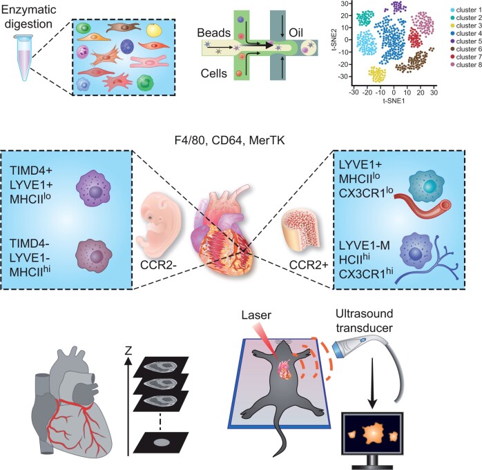
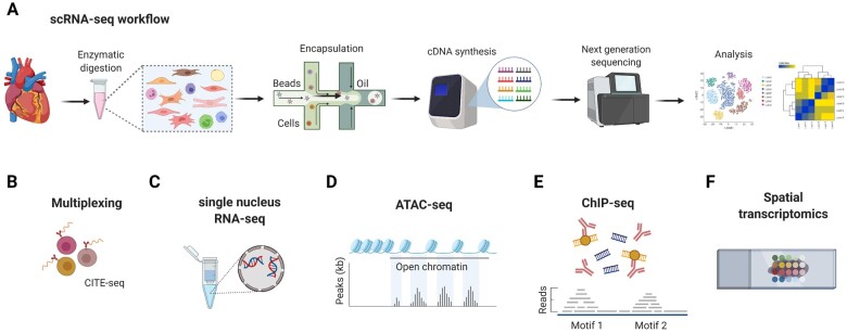
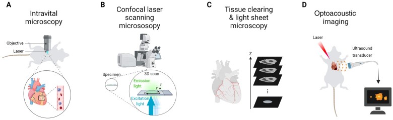
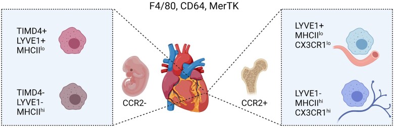
The increasing use of single-cell immune profiling and advanced microscopic imaging technologies has deepened our understanding of the cardiac immune system, confirming that the heart contains a broad repertoire of innate and adaptive immune cells. Leucocytes found in the healthy heart participate in essential functions to preserve cardiac homeostasis, not only by defending against pathogens but also by maintaining normal organ function. In pathophysiological conditions, cardiac inflammation is implicated in healing responses after ischaemic or non-ischaemic cardiac injury. The aim of this review is to provide a concise overview of novel methodological advancements to the non-expert readership and summarize novel findings on immune cell heterogeneity and functions in cardiac disease with a focus on myocardial infarction as a prototypic example. In addition, we will briefly discuss how biological sex modulate the cardiac immune response. Finally, we will highlight emerging concepts for novel therapeutic applications, such as targeting immunometabolism and nanomedicine.

Keywords: Bioimaging; Heart failure; Immunometabolism; Nanomedicine; Sexual dimorphism; Single-cell RNA sequencing.


Figures

Graphical Abstract
Graphical Abstract
State of the art technologies to study cardiac immunology include single cell RNA sequencing and advanced microscopic imaging, revealing an unprecedented immune cell heterogeneity of the heart.
Figure 1
Figure 1
Overview of scRNA-seq and related technological advancements to study the cardiac transcriptome. (A) The technologies used for scRNA-seq mostly employ microfluidics platforms for single-cell encapsulation as initial step of sample preparation, combined with next-generation sequencing approaches for entire transcriptome measurements. (B) To improve the precision of immune cell subset identification, which might be relevant for rare cell populations, oligonucleotide-barcoded antibody labelling, or lipid-tagged indices for barcoding of cells prior to their processing for scRNA-seq can be used.  ,   This multimodal measurement of cell surface labels in combination with transcript expression allows combining several biological samples in one scRNA-seq library, which is referred to as multiplexing. This reduces inter-sample technical bias and facilitates exclusion of cellular doublets, thereby limiting analytical artefacts. (C) An alternative to scRNA-seq is the single nucleus RNA-seq (snRNA-seq) method, which might be preferable when working with tissues that are difficult to dissociate or frozen and large cells such as cardiomyocytes.  ,   Other technical advancements involve combined methods for epigenetic profiling with scRNA-seq, such as (D) assay for transposase-accessible chromatin using sequencing (ATAC-seq) or (E) chromatin immunoprecipitation followed by sequencing (ChIP–seq). ATAC-seq allows sequencing of the regions of the genome with open or accessible chromatin, while ChIP-seq enables genome-wide profiling of DNA-binding proteins and histone modifications. (F) Novel developments combine transcriptomics with spatial information, in order to overcome the issue that the information on cellular distribution gets lost due to the generation of single-cell suspensions.
Figure 2
Figure 2
Overview of advanced imaging technologies. (A) Intravital microscopy of the beating mouse heart is an invasive method that involves thoracic surgery and cardiac tissue stabilizers. In addition, it requires acquisition gating algorithms to avoid moving artefacts caused by the cardiac and respiratory cycles. Due to these technical requirements and need for highly specialized knowledge, the methodology has not been broadly applied to study immune cell behaviour in cardiac disease models. (B) Confocal laser scanning microscopy allows scanning multiple layers of a sample and subsequent 3-dimensional image reconstruction. (C) For tissue clearing, the heart is processed by organic solvent- or aqueous-based clearing or hydrogel embedding tissue clearing. When combined with diverse labelling methods (such as immunolabelling or use of reporter mice) and high-throughput optical sectioning light sheet microscopy, tissue clearing enables whole-body and whole-organ imaging at cellular or subcellular resolution. (D) Optoacoustic imaging is a technology that uses ultrasound sensors to visualize structures based on the optoacoustic effect, which refers to the generation of acoustic waves from endogenous chromophores in biological tissues (e.g. haem) following pulsed-light illumination. The light absorption by these endogenous components results in thermoelastic expansion of the tissue and generation of mechanical waves, which are resistant to scattering, thereby enabling higher spatial resolution and greater penetration depths than emitted light in optical imaging. In addition to label-free imaging of endogenous structures such as blood vessels, optoacoustic imaging can also be used to track transplanted immune cells in entire living animals, which is achieved by near-infrared fluorescent optoacoustic probes.
Figure 3
Figure 3
Cardiac resident macrophage heterogeneity during steady state. Macrophages in the healthy adult heart are highly heterogeneous, which is related to their distinct ontogeny (embryonic vs. adult bone marrow monocyte progenitor-derived) and tissue microenvironment. A common set of markers to identify macrophages includes F4/80, CD64, and MerTK. CCR2 macrophages largely originate from embryonic (yolk sac and foetal liver) origin and can be further subdivided into LYVE1+ MHCIIlo and LYVE1 MHCIIhi. TIMD4 recently emerged as additional marker of CCR2 MHCIIlo resident macrophages that is fully maintained independent of blood monocytes, whereas CCR2 MHCIIhi TIMD4 resident macrophages are partially replaced. CCR2+ macrophages are derived from bone marrow haematopoiesis, giving raise to CCR2+ blood monocytes that enter the adult heart. Another dimension of cardiac bone marrow monocyte-derived macrophage heterogeneity involves the separation into LYVE1+ MHCIIlo CX3CR1lo and LYVE1 MHCIIhi CX3CR1hi tissue macrophage subsets with distinct localization and functional specializations.
Figure 4
Figure 4
New insights into immune cell functions and phenotypes in ischaemic cardiac injury. (1) Cardiac macrophages in the healthy heart are mostly CCR2 and of foetal origin, as opposed to monocyte-derived CCR2 expressing macrophages, which infiltrate the heart after MI. Ischaemic injury reduces CCR2 TIMD4+ and TIMD4 resident macrophage abundance, whereas CCR2+ monocyte-derived macrophages expand and adopt heterogeneous phenotypes within infarcted tissue. (2) The cGAS-STING signalling pathway is activated in CCR2+ macrophages by extracellular DNA released from dying cells, thereby promoting an IRF3-dependent type I IFN release and monocyte recruitment. (3) Engulfment of dying cells by macrophages within the infarcted myocardium results in elevated macrophage fatty acid content, which stimulates mitochondrial respiration. This metabolic signalling pathway promotes an anti-inflammatory macrophage response during wound healing after MI. (4) The different post-MI healing stages are also accompanied by diverse neutrophil subsets, with an aged neutrophil phenotype accumulating in the circulation over time. Conversely, heart infiltrating neutrophils acquire a high expression level of the surface lectin SiglecF, resembling an activated phenotype previously observed in cancer. (5) Basophil-derived IL4 and IL14 production plays an essential role in the infarcted heart, by balancing the ratio of proinflammatory monocytes to reparative Ly6Clo macrophages. (6) A new macrophage phenotype expressing GATA6 has been identified in the pericardial cavity fluid, which is recruited to the heart in response to MI and plays a protective role by preventing excessive cardiac fibrosis. (7) The pericardial adipose tissue contains lymphoid clusters [fat-associated lymphoid clusters (FALCs)], which expand after MI. Activated dendritic cells migrate from the infarcted myocardium to pericardial adipose tissue FALCs and induce B and T cell proliferation. (8) Moreover, an increasing number of patrolling Ly6Clo monocytes, which express PDL1, interacts with T cells within pericardial FALCs and induces their apoptosis, possibly as an inhibitory mechanism to prevent sustained lymphocyte activation. (9) IL2 promotes ILC2 expansion in the circulation and the pericardial adipose tissue after MI, and is associated with an improved cardiac function, while genetic depletion of ILC2s worsened cardiac recovery post-MI. (10) An innate-like B cell subset within pericardial FALCs expresses high amounts of GM-CSF in response to MI, which induces IL23 and IL17 cytokine release by other pericardial immune cells. (11) The systemic cytokine increase triggers the production of the granulocytic factor G-CSF, which, in turn promotes de novo production of neutrophils in the bone marrow, thereby maintaining the neutrophil supply to the ischaemic heart.

References

    1. Swirski FK, Nahrendorf M. Cardioimmunology: the immune system in cardiac homeostasis and disease. Nat Rev Immunol 2018;18:733–744. - PubMed
    1. Marelli-Berg FM, Aksentijevic D. Immunometabolic cross-talk in the inflamed heart. Cell Stress 2019;3:240–266. - PMC - PubMed
    1. Jessup M, Brozena S. Heart failure. N Engl J Med 2003;348:2007–2018. - PubMed
    1. Lam CSP, Arnott C, Beale AL et al. Sex differences in heart failure. Eur Heart J 2019;40:3859c–3868c. - PubMed
    1. Klein SL, Flanagan KL. Sex differences in immune responses. Nat Rev Immunol 2016;16:626–638. - PubMed

Publication types