

Restoring Rotation Center in Total Hip Arthroplasty for Developmental Dysplasia of the Hip with the Assistance of Three Dimensional Printing Technology: A Pilot Study
- PMID: 34898037
- PMCID: PMC8755880
- DOI: 10.1111/os.13183
Restoring Rotation Center in Total Hip Arthroplasty for Developmental Dysplasia of the Hip with the Assistance of Three Dimensional Printing Technology: A Pilot Study
Abstract
Objective: To develop a new method to restore hip rotation center exactly and rapidly in total hip arthroplasty (THA) with the assistance of three dimensional (3D) printing technology and evaluate its clinical and radiological outcomes.
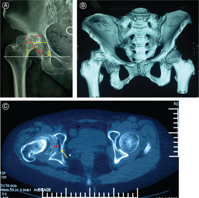
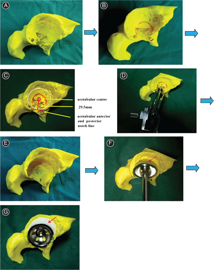
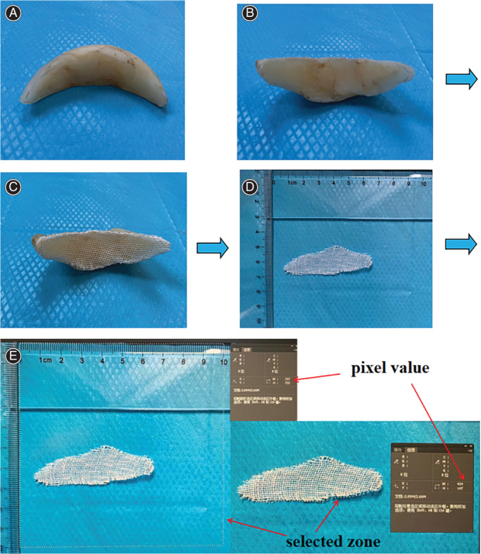
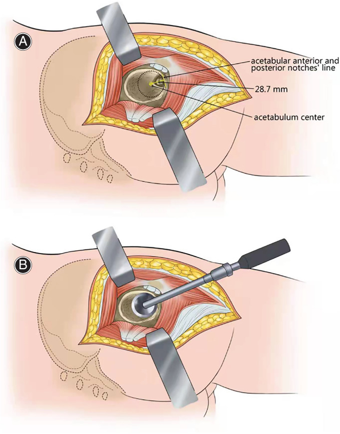
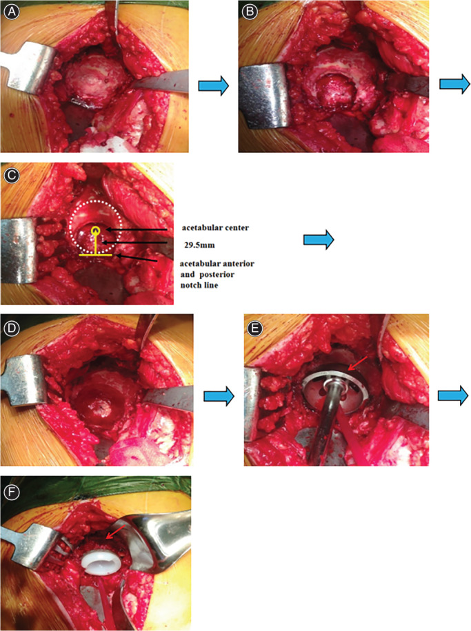
Methods: From March 2014 to July 2018, a total of 17 patients (five hips of four men and 16 hips of 13 women) with end-stage osteoarthritis secondary to developmental dysplasia of the hip who underwent THA were analyzed and followed up retrospectively. The average age is 58.00 ± 8.12 years (range from 45 to 71 years). Simulated operations were performed on 3D printed hip models for preoperative planning. The morphology of Harris fossa and acetabular notches were recognized and restored to locate the acetabular center. The size of bone defect was measured by the bone wax method. The agreement on the size of acetabular cup and bone defect between simulated operations and actual operations were analyzed. Harris Hip Score (HHS) was used to evaluate the recovery of hip joint function. The vertical distance and horizontal distance of the rotation center on the pelvis plain radiograph were measured, which were used to assess the efficacy of restoring hip rotation center and acetabular cup migration.
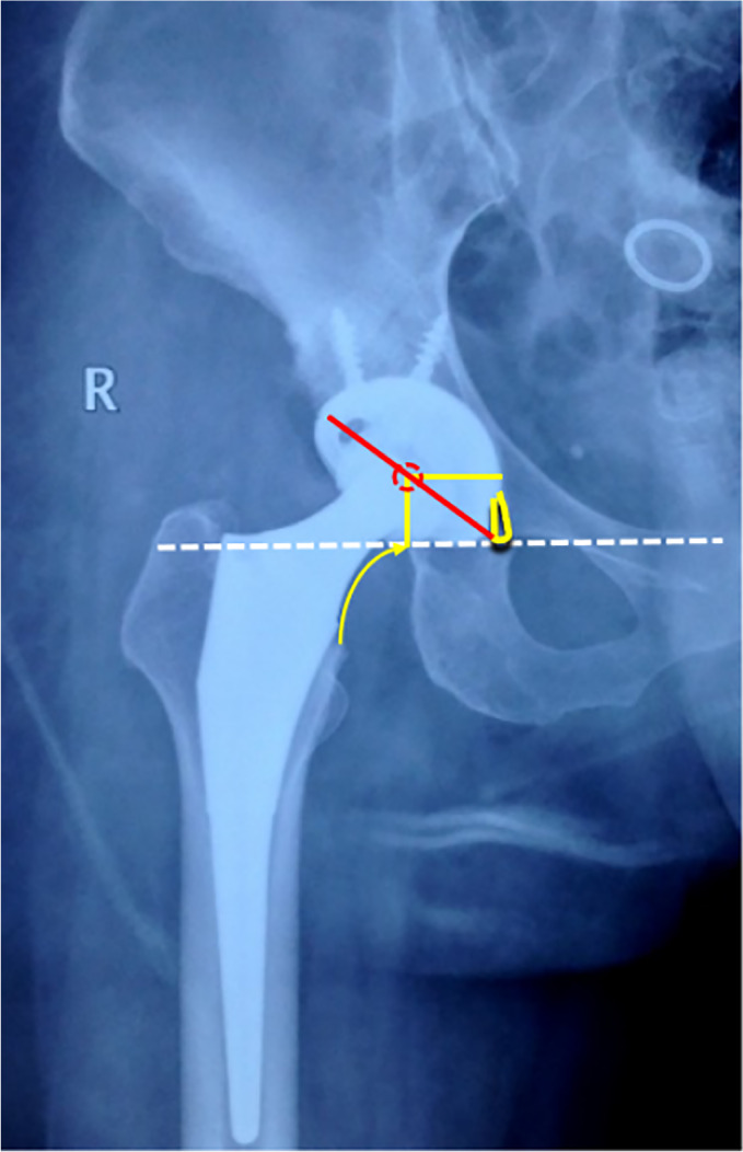
Results: The mean sizes of bone defect in simulated operations and THA were 4.58 ± 2.47 cm2 and 4.55 ± 2.57 cm2 respectively. There was no significant difference statistically between the sizes of bone defect in simulated operations and the actual sizes of bone defect in THA (t = 0.03, P = 0.97). The sizes of the acetabular cup of simulated operations on 3D print models showed a high rate of coincidence with the actual sizes in the operations (ICC = 0.93). All 17 patients were available for clinical and radiological follow-up. The average follow-up time was 18.35 ± 6.86 months (range, 12-36 months. The average HHS of the patients was improved from (38.33 ± 6.07) preoperatively to the last follow-up (88.61 ± 3.44) postoperatively. The mean vertical and horizontal distances of hip rotation center on the pelvic radiographs were restored to 15.12 ± 1.25 mm and 32.49 ± 2.83 mm respectively. No case presented dislocation or radiological signs of loosening until last follow-up.
Conclusions: The application of 3D printing technology facilitates orthopedists to recognize the morphology of Harris fossa and acetabular notches, locate the acetabular center and restore the hip rotation center rapidly and accurately.
Keywords: 3D printing; Arthroplasty; Developmental dysplasia of the hip; Hip; Rotation center.
© 2021 The Authors. Orthopaedic Surgery published by Chinese Orthopaedic Association and John Wiley & Sons Australia, Ltd.
Figures
References
-
- Chen M, Luo ZL, Wu KR, Zhang XQ, Ling XD, Shang XF. Cementless total hip arthroplasty with a high hip center for hartofilakidis type B developmental dysplasia of the hip: results of midterm follow‐up. J Arthroplasty, 2016, 31: 1027–1034. - PubMed
-
- Yang TC, Chen CF, Tsai SW, Chen WM, Chang MC. Does restoration of hip center with subtrochanteric osteotomy provide preferable outcome for Crowe type III‐IV irreducible development dysplasia of the hip. J Chin Med Assoc, 2017, 80: 803–807. - PubMed
-
- Nie Y, Pei F, Shen B, Kang P, Li Z. Implication of acetabular width on the anteroposterior pelvic radiograph of patients with developmental dysplasia of the hip. J Arthroplasty, 2015, 30: 489–494. - PubMed
-
- Hardt S, Hube R, Perka C. Total hip arthroplasty for high hip dislocation. Z Orthop Unfall, 2020, 158: 170–183. - PubMed
MeSH terms
Grants and funding
LinkOut - more resources
Full Text Sources
Medical
