. 2022 Jan 21;11(1):61-68.
doi: 10.1021/acssynbio.1c00398. Epub 2021 Dec 13.

Analysis of Three Architectures for Controlling PTP1B with Light

Affiliations

Analysis of Three Architectures for Controlling PTP1B with Light

Akarawin Hongdusit et al. ACS Synth Biol. .

Abstract

Photosensory domains are powerful tools for placing proteins under optical control, but their integration into light-sensitive chimeras is often challenging. Many designs require structural iterations, and direct comparisons of alternative approaches are rare. This study uses protein tyrosine phosphatase 1B (PTP1B), an influential regulatory enzyme, to compare three architectures for controlling PTPs with light: a protein fusion, an insertion chimera, and a split construct. All three designs permitted optical control of PTP1B activity in vitro (i.e., kinetic assays of purified enzyme) and in mammalian cells; photoresponses measured under both conditions, while different in magnitude, were linearly correlated. The fusion- and insertion-based architectures exhibited the highest dynamic range and maintained native localization patterns in mammalian cells. A single insertion architecture enabled optical control of both PTP1B and TCPTP, but not SHP2, where the analogous chimera was active but not photoswitchable. Findings suggest that PTPs are highly tolerant of domain insertions and support the use of in vitro screens to evaluate different optogenetic designs.

Keywords: light-oxygen-voltage domains; optogenetics; protein engineering; protein tyrosine phosphatases; signaling.


Conflict of interest statement

The authors declare no competing financial interest.

Figures

Figure 1.
Figure 1.
Development of alternative optogenetic architectures for PTP1B. (A) A depiction of PTP1BPS. In this fusion, the A′α helix of LOV2 is connected to the allosterically influential α7 helix of PTP1B. Illumination of LOV2 with blue light causes its terminal helices to unwind, destabilizes the α7 helix of PTP1B, and disrupts catalytic activity. (B) The design concept for an “insertion” chimera: LOV2 is inserted within a flexible loop on the surface of PTP1B; illumination of LOV2 causes structural distortions in PTP1B that disrupt its catalytic activity. (C) The design concept for a “split” construct: Two halves of PTP1B are held together by a noncovalent LOV2-Zdk1 complex; illumination of LOV2 causes the heterodimeric complex to dissociate—or to undergo a conformational change caused by the dissociation of LOV2-Zdk1—and disrupts catalytic activity. (D) The dark-state catalytic efficiency (CEdark = [kcat/KM]dark) and the dynamic range (DR = [kcat/KM]dark/[kcat/KM]light) of various constructs. Constructs with an asterisk were either not active or not expressible in E. coli. (E,F) Initial rates of PTP-catalyzed hydrolysis of p-nitrophenyl phosphate (pNPP) by high-DR variants of the (E) insertion and (F) split constructs in the presence and absence of light (455 nm). Error bars show standard error of the mean (n = 3 independently prepared reactions). Table S1 provides source data.
Figure 2.
Figure 2.
Analysis of natively localized constructs. (A) Images of COS-7 cells expressing RFP-tagged variants of PTP1B: PTP1B435 is the wild-type protein, which contains a C-terminal region (a disordered proline-rich domain followed by a short membrane anchor) that localizes it to the endoplasmic reticulum (ER). PTP1BPS* is a version of PTP1BPS that includes residues 299–435 from the wild-type enzyme attached to its C-terminus; I1* and S1A* include the same region of the full-length enzyme. PTP1B435, PTP1BPS*, and I1* exhibit indistinguishable localization patterns that are consistent with localization to the ER; for S1A*, fluorescence in the cytosol suggests that a significant fraction of its N-terminal half (i.e., RFP-PTP1B-Zdk1) is dissociated from its C-terminal half (i.e., ER-localized LOV2-PTP1B; scale bar, 10 μm). Additional images appear in Figure S3. (B,C) ELISA-based measurements of IR phosphorylation in HEK293T/17 stably expressing variants of PTP1B after 10 min exposure to light (455 nm) or darkness. The DR (i.e., the ratio of light- to dark-state signals) appears above the bars for each construct. (B) Illumination increases IR phosphorylation for all constructs. (C) The introduction of LOV2-stabilizing mutations does not improve the dynamic range of S1A*. The plotted data depict the mean and SE for measurements of n = 3 biological replicates. The error in DR depicts propagated SE for n = 3 biological replicates under each condition (light and dark). Source data appear in Table S6.
Figure 3.
Figure 3.
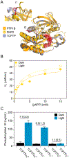
Extension of the insertion-based approach to other PTPs. (A) Aligned crystal structures of the catalytic domains of PTP1B, SHP2, and TCPTP (pdb entries 2cm2, 3zm1, and 1l8k, respectively). Highlights: competitive inhibitor (red spheres, pdb entry 2f71), insertion site I1 (PTP1B205–206, TCPTP206–207, and SHP2449–450; red sticks), and insertion site I2 (PTP1B186–187, red sticks). (B) Initial rates of SHP2I1-catalyzed hydrolysis of pNPP in the presence and absence of light (455 nm). Values of kcat and KM are indistinguishable (p < 0.05). (C) ELISA-based measurements of IR phosphorylation in HEK293T/17 cells stably expressing photoswitchable variants of TCPTP after 10 min exposure to light (455 nm) or darkness. TCPTPPS* is a version of TCPTPPS that includes residues 318–415 from the wild-type enzyme (i.e., the ER anchor) attached to its C-terminus; TCPTPI1* and TCPTPI2* include the same region of the full-length enzyme. Illumination increases IR phosphorylation for TCPTPPS* and TCPTPI1*, but not TCPTPI2*. The plotted data depict the mean and SE for measurements of n = 3 biological replicates. The DR appears above the bars for each construct; here, error depicts propagated SE for n = 3 biological replicates under each condition (light and dark). Tables S1 and S6 provide source data.

References

    1. Toettcher JE; Weiner OD; Lim WA Using optogenetics to interrogate the dynamic control of signal transmission by the Ras/Erk module. Cell 2013, 155, 1422–1434. - PMC - PubMed
    1. Van Haren J; Charafeddine RA; Ettinger A; Wang H; Hahn KM; Wittmann T Local control of intracellular microtubule dynamics by EB1 photodissociation. Nat. Cell Biol 2018, 20, 252–261. - PMC - PubMed
    1. Kim N; Kim JM; Lee M; Kim CY; Chang KY; Heo W. Do. Spatiotemporal control of fibroblast growth factor receptor signals by blue light. Chem. Biol 2014, 21, 903–912. - PubMed
    1. Zhou XX; Fan LZ; Li P; Shen K; Lin MZ Optical control of cell signaling by single-chain photoswitchable kinases. Science (Washington, DC, U. S.) 2017, 355, 836–842. - PMC - PubMed
    1. Bantscheff M; Eberhard D; Abraham Y; Bastuck S; Boesche M; Hobson S; Mathieson T; Perrin J; Raida M; Rau C; Reader V; Sweetman G; Bauer A; Bouwmeester T; Hopf C; Kruse U; Neubauer G; Ramsden N; Rick J; Kuster B; Drewes G Quantitative chemical proteomics reveals mechanisms of action of clinical ABL kinase inhibitors. Nat. Biotechnol 2007, 25, 1035–1044. - PubMed

Publication types