. 2022 Feb 15;204(2):e0043221.
doi: 10.1128/JB.00432-21. Epub 2021 Dec 13.

Distinct Interaction Mechanism of RNA Polymerase and ResD at Proximal and Distal Subsites for Transcription Activation of Nitrite Reductase in Bacillus subtilis

Affiliations

Distinct Interaction Mechanism of RNA Polymerase and ResD at Proximal and Distal Subsites for Transcription Activation of Nitrite Reductase in Bacillus subtilis

Hannah Jacob et al. J Bacteriol. .

Abstract

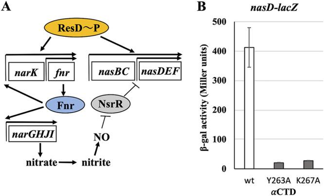
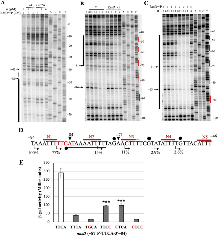
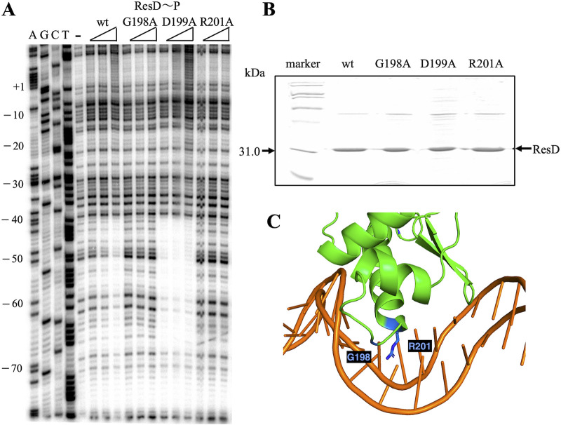
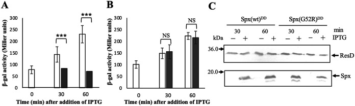
The ResD-ResE signal transduction system plays a pivotal role in anaerobic nitrate respiration in Bacillus subtilis. The nasD operon encoding nitrite reductase is essential for nitrate respiration and is tightly controlled by the ResD response regulator. To understand the mechanism of ResD-dependent transcription activation of the nasD operon, we explored ResD-RNA polymerase (RNAP), ResD-DNA, and RNAP-DNA interactions required for nasD transcription. Full transcriptional activation requires the upstream promoter region where five molecules of ResD bind. The distal ResD-binding subsite at -87 to -84 partially overlaps a sequence similar to the consensus distal subsite of the upstream (UP) element with which the Escherichia coli C-terminal domain of the α subunit (αCTD) of RNAP interacts to stimulate transcription. We propose that interaction between αCTD and ResD at the promoter-distal site is essential for stimulating nasD transcription. Although nasD has an extended -10 promoter, it lacks a reasonable -35 element. Genetic analysis and structural simulations predicted that the absence of the -35 element might be compensated by interactions between σA and αCTD and between αCTD and ResD at the promoter-proximal ResD-binding subsite. Thus, our work suggested that ResD participates in nasD transcription activation by binding to two αCTD subunits at the proximal and distal promoter sites, representing a unique configuration for transcription activation. IMPORTANCE A significant number of ResD-controlled genes have been identified, and transcription regulatory pathways in which ResD participates have emerged. Nevertheless, the mechanism of how ResD activates transcription of different genes in a nucleotide sequence-specific manner has been less explored. This study suggested that among the five ResD-binding subsites in the promoter of the nasD operon, the promoter-proximal and -distal ResD-binding subsites play important roles in nasD activation by adapting different modes of protein-protein and protein-DNA interactions. The finding of a new type of protein-promoter architecture provides insight into the understanding of transcription activation mechanisms controlled by transcription factors, including the ubiquitous response regulators of two-component regulatory systems, particularly in Gram-positive bacteria.

Keywords: Bacillus subtilis; RNA polymerase subunits; ResD-dependent transcription; UP element; nitrite reductase.


Conflict of interest statement

The authors declare no conflict of interest.

Figures

FIG 1
FIG 1
ResD-dependent activation of nasD transcription. (A) A schematic view of regulatory cascade involved in nasD transcriptional control. Arrowheads and perpendicular ends show positive and negative effects, respectively. Transcription regulators are marked in color. (B) Effect of K267A and Y263A of α on ResD-dependent nasD transcription under nitrate respiratory conditions. Transcriptional fusion of the nasD promoter (–93 to +257) to lacZ was used for measurement of β-galactosidase (β-gal) activity using cultures at T0 (the end of exponential growth). Averages and standard deviations representative of three biological replicates are shown.
FIG 2
FIG 2
Promoter-distal ResD-αCTD interaction subsite is required for stimulation of nasD transcription. (A to C) The labeled-coding strand of the nasD (−138 to +22) promoter was used for binding reactions with ResD phosphorylated by ResE (ResD∼P) and/or full-length α. Nucleotide positions of the sequence ladders are marked relative to the transcription start site (+1). (A) Protected region in the presence of both ResD∼P and wild-type α is marked with a black bar and a dashed line. (B) Region protected by ResD∼P is marked with a black bar; −84 was protected by α and weakly by ResD∼P. (C) A region marked with a black bar was protected by the presence of ResD∼P and α. Red lines next to sequencing ladders in panels B and C indicate protected regions by previous hydroxyl radical footprinting by ResD∼P (10). (D) nasD transcription requires the upstream region of the nasD promoter. Red lines with N1 to N5 above the nasD sequence are residues protected from hydroxyl radical attack by ResD∼P binding (10). The percentages under the nucleotide sequence show nasD-lacZ activation relative to the longest promoter (−93 to +257) fused to lacZ, and the arrows denote the 5′ end of deletions (12). Three G residues are marked by black circles above the sequence, and −87 5′-TTCA-3′ −84, labeled in red, is identical to the sequence previously reported as the ResD-binding site at fnr (30). The sequence between −84 and −75 is identical or similar to the consensus distal UP element sequences (31, 32). (E) Effect of base substitutions of nasD (−87 to −84) on nasD transcription. The wild-type and mutant strains at T0 were used for the measurement of β-galactosidase activity. Averages and standard deviations from three biological replicates are shown. Statistical differences indicated by asterisks were compared to the wild type. ***, P < 0.001.
FIG 3
FIG 3
α-Loop of ResDc participates in DNA binding. (A) DNase I footprinting of nasD (−138 to +22) with the wild-type (wt) and mutant ResD∼P. The labeled coding strand was used with 1, 2, and 4 μM ResD∼P. Amino acid substitutions in the ResD α loop are indicated on top. Nucleotide positions of the sequence ladder are marked relative to the transcription start site (+1). (B) ResD was overexpressed and purified from E. coli, and 5 μg total protein was used for Western blotting to confirm ResD stability and for use in DNase I footprinting. (C) Predicted structural model for ResD-DNA interaction using i-TASSER.
FIG 4
FIG 4
Overexpression of Spx decreases nasD transcription without affecting ResD protein levels. (A) Effect of Spx(wt)DD overexpression on nasD-lacZ transcription. When cultures reached an OD600 of 0.1 to 0.2, 100 μM IPTG was added. Cultures were further incubated with IPTG (filled box) or without IPTG (open box). Cells were harvested 30 and 60 min after treatment with IPTG, and nasD-lacZ activity was measured. The activity is shown as averages from three biological replicates with standard deviations. Statistical differences are indicated by asterisks. *, P < 0.05; **, P < 0.01; ***, P < 0.001; NS, not significant. (B) nasD-lacZ activity was measured as described for panel A, except that SpxG52RDD, which is unable to interact with αCTD, was used (27, 28, 41). (C) B. subtilis cells were harvested at the same time as that shown in panels A and B to assess protein levels by Western blotting. A nitrocellulose membrane was cut in half after transferring proteins, and each membrane was used for the detection of ResD or Spx. The blot is a representative of three biological samples.
FIG 5
FIG 5
Effect of amino acid substitutions of ResDc N-terminal β-sheet on nasD transcription. (A) Amino acid sequence alignment of ResD (residues 141 to 171) and Spx (residues 32 to 67). G52, the residue of Spx required for the interaction with Y263 of αCTD, is marked in red. Residues conserved between ResD and Spx are marked. A plus sign indicates similar residues, and a minus sign shows residues absent from ResD. (B) Cells were grown under nitrate respiratory conditions in the presence of 1 mM IPTG. The ratio of mutant to wild-type expression levels is from samples taken at T0 (the end of exponential growth). White bars represent significantly affecting residue substitutions, while black bars represent residues not significantly affecting expression. Each experiment was repeated using at least three biological replicates, and the data points are shown as the average ratio of mutant to wild type. Statistical differences are indicated by asterisks. *, P < 0.05; **, P < 0.01; ***, P < 0.001; P values were compared to the wild type. (C) Western blot analysis of ResD proteins. Cells harvested at the same time as those used for measuring nasD-lacZ in panel B were disrupted by bead beating, and 5 μg of protein was applied to 12% SDS polyacrylamide gels. Only three strains (wt, R153A, and D148A) were examined with and without IPTG, and other samples were obtained from the strains induced by IPTG. Positions of molecular mass markers (30 kDa) are shown on the left. Mutant strains in blue showed moderately but significantly reduced nasD transcription and ResD concentration similar to that of the wild-type strain. ResD mutants marked with purple are unstable and unable to activate transcription. ResDH152A protein is stable but defective in transcription activity. (D to G) Models for ResDc-αCTD interaction predicted using the PyDock and Haddock servers. The interacting residues of ResDc and αCTD are shown in green and cyan, respectively, with the red dashes representing hydrogen bonds. The interacting residues per the top-scoring PyDock predicted model are ResDH152-αY263, ResDA151-αN264 (D), and (E) ResDT164-αD257. The interacting residues per the top-scoring Haddock predicted model are ResDH152-αK267, ResDD148-αR268, and ResDN138-αR268 (F) and ResDR153-αN264 and ResDA151-αY263 (G). (H) A space-filling model of ResDc showing residues that interact with the surface including αY263. (I) A space-filling model of αCTD showing the surface patch including αY263.
FIG 6
FIG 6
Region 4 of σA is required for nasD activation. (A) Effects of amino acid substitutions of σA on nasD-lacZ and narG-lacZ. Cells were cultured under nitrate respiratory conditions, and samples were taken at T0. The data shown are averages from at least three biological replicates with standard deviations. (B) σA H359R, K356E, and R362A, which reduced nasD transcription, were further examined to determine whether the addition of KNO2 restores transcription. Cells were cultured under fermentative conditions (2× YT with 0.5% glucose and 0.5% pyruvate). When the OD600 reached 0.1 to 0.2, KNO2 (final concentration, 5 mM) was added to one set of cultures. Cells cultured with and without KNO2 were harvested after 30 min and 60 min. Statistical differences are indicated by asterisks. **, P < 0.01; ***, P < 0.001; P values in panel A were compared to the wild type.
FIG 7
FIG 7
Model for activation of the nasD promoter by ResD. Proposed locations of σA, αCTD, and ResDc on nasD are marked with dashed circles. Red lines in N1 to N5 above the nasD sequence showed protection from hydroxyl radical attack by ResD binding (10). The extended −10 (TG − 10) and the −35 region are shown by using red color for residues that match the canonical −10 (TATAAT) and −35 (TTGACA) sequences. The −87 5′-TTCA-3′ −84 sequence is marked with blue, and the adjacent UP element is lined in blue. ResD1 with an arrow is proposed to bind to the opposite face of the promoter DNA as αCTD and σA.

Similar articles

Cited by

References

    1. Schau M, Eldakak A, Hulett FM. 2004. Terminal oxidases are essential to bypass the requirement for ResD for full Pho induction in Bacillus subtilis. J Bacteriol 186:8424–8432. 10.1128/JB.186.24.8424-8432.2004. - DOI - PMC - PubMed
    1. Sun G, Birkey SM, Hulett FM. 1996. Three two-component signal-transduction systems interact for Pho regulation in Bacillus subtilis. Mol Microbiol 19:942–948. - PubMed
    1. Zhang X, Hulett FM. 2000. ResD signal transduction regulator of aerobic respiration in Bacillus subtilis; cta promoter regulation. Mol Microbiol 37:1208–1219. 10.1046/j.1365-2958.2000.02076.x. - DOI - PubMed
    1. Nakano MM, Zuber P. 1998. Anaerobic growth of a “strict aerobe” (Bacillus subtilis). Annu Rev Microbiol 52:165–190. 10.1146/annurev.micro.52.1.165. - DOI - PubMed
    1. Nakano MM, Zuber P. 2002. Anaerobiosis, p 393–404. In Sonenshein AL, Hoch JA, Losick R (ed), Bacillus subtilis and its closest relatives: from genes to cells. ASM Press, Washington, DC.

Publication types

MeSH terms

LinkOut - more resources