Review
. 2022 Jan 1;102(1):411-454.
doi: 10.1152/physrev.00002.2021.

The evolution of regulated cell death pathways in animals and their evasion by pathogens

Affiliations
Review

The evolution of regulated cell death pathways in animals and their evasion by pathogens

Bart Tummers et al. Physiol Rev. .

Abstract

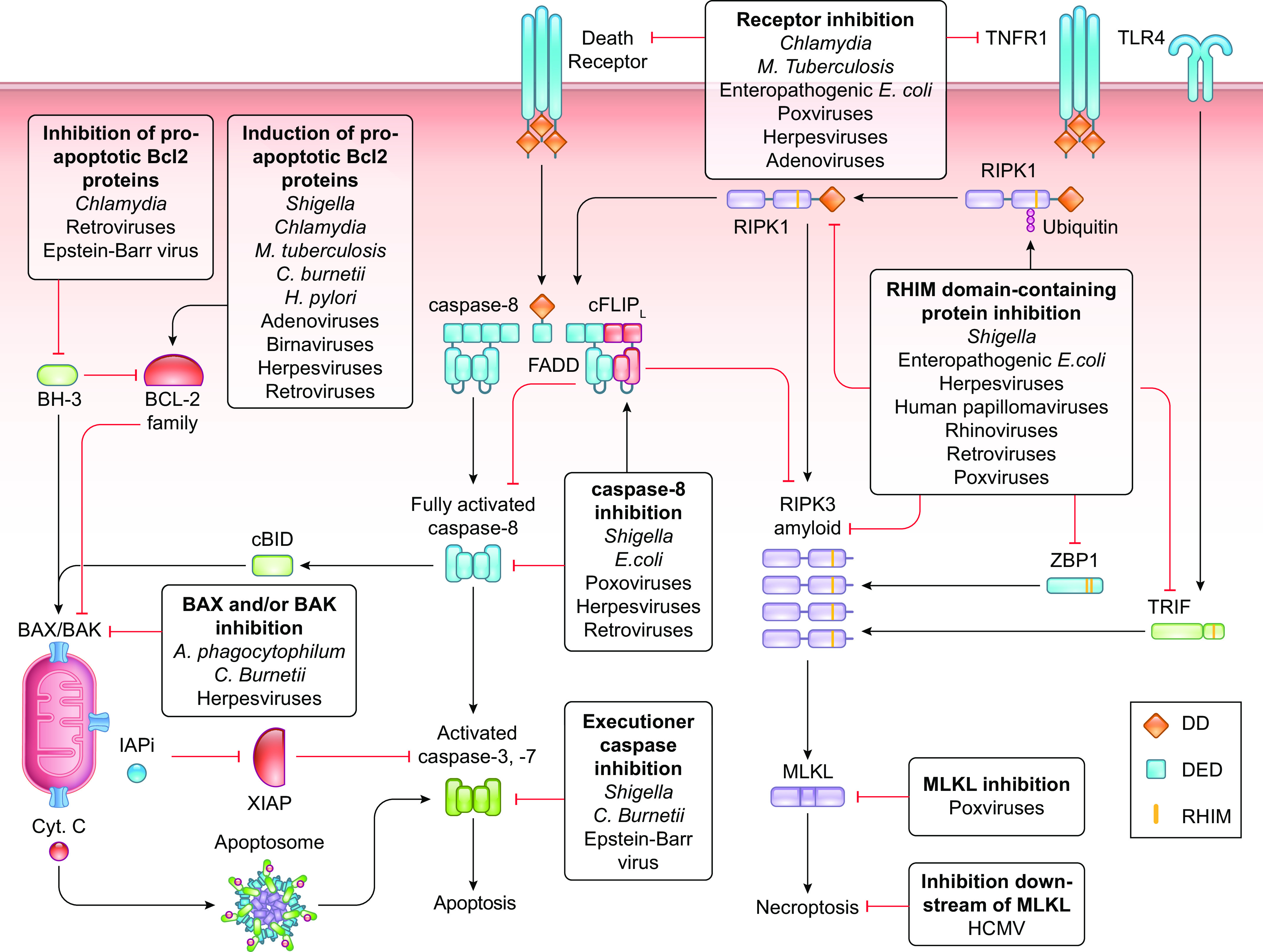
The coevolution of host-pathogen interactions underlies many human physiological traits associated with protection from or susceptibility to infections. Among the mechanisms that animals utilize to control infections are the regulated cell death pathways of pyroptosis, apoptosis, and necroptosis. Over the course of evolution these pathways have become intricate and complex, coevolving with microbes that infect animal hosts. Microbes, in turn, have evolved strategies to interfere with the pathways of regulated cell death to avoid eradication by the host. Here, we present an overview of the mechanisms of regulated cell death in Animalia and the strategies devised by pathogens to interfere with these processes. We review the molecular pathways of regulated cell death, their roles in infection, and how they are perturbed by viruses and bacteria, providing insights into the coevolution of host-pathogen interactions and cell death pathways.

Keywords: apoptosis; cell death; infection; necroptosis; pyroptosis.


Conflict of interest statement

D.R.G. consults for Ventus Therapeutics and Inzen Therapeutics. B.T. has no conflicts of interest, financial or otherwise, to disclose.

Figures

None
Graphical abstract
FIGURE 1.
FIGURE 1.
Domain structure of the caspases discussed here. Schematic representation of initiator caspases-2, -9, -8, and -10 and DRONC, executioner caspases-3, -6, and -7, DRICE, and CED-3, inflammatory caspases-1, -4, -5, -11, and -12, caspase-14 (involved in cellular differentiation), a subset of caspases present in Hydra, and one of the pyrin domain (PYD)-containing caspases present in zebrafish. a.a., Amino acids; CARD, caspase-recruitment domain; Ce, Caenorhabditis elegans; DD, death domain; DED, death effector domain; Dm, Drosophila melanogaster; Dr, Dania rerio; H, Hydra spp.; Hs, Homo sapiens; Mm, Mus musculus; ?, prodomain of unknown structure.
FIGURE 2.
FIGURE 2.
Pathways of pyroptosis. A: the noncanonical inflammasome is activated upon recognition of LPS by the inflammatory caspases-4, -5, and -11, resulting in dimerization, activation, and processing of gasdermin D (GSDMD). Activated GSDMD induces plasma membrane pore formation by oligomerization to execute pyroptosis. B: the canonical inflammasome is activated upon engagement of inflammasome-inducing receptors, which recruit the inflammatory caspase-1, either directly via caspase-recruitment domain (CARD) interactions or indirectly via pyrin domain (PYD) and CARD interactions mediated by the adaptor protein ASC, forming the inflammasome. Activated caspase-1 cleaves pro-IL-1β and pro-IL-18 into their biologically active forms and activates GSDMD to execute pyroptosis.
FIGURE 3.
FIGURE 3.
The caspase-9 pathway of apoptosis. Comparison between the caspase-9 pathway of apoptosis in most animals, arthropods, and nematodes. A: in most animals, the caspase-9 pathway of apoptosis is mediated by the BCL-2 effector proteins BAX and BAK to induce mitochondrial outer membrane permeabilization (MOMP). Activation of BCL-2 effector proteins is regulated by the balance between proapoptotic and antiapoptotic Bcl-2 proteins. MOMP releases cytochrome c and inhibitor of apoptosis (IAP) inhibitors (IAPi) into the cytosol, where cytochrome c binds and activates Apoptotic Protease Activating Factor 1 (APAF-1) to recruit the initiator caspase-9, forming the apoptosome. Activated caspase-9 cleaves and activates the executioner caspases-3 and -7 to mediate apoptosis. IAPi released by MOMP block the suppressive effects of the caspase inhibitor X-linked inhibitor of apoptosis protein (XIAP), allowing the caspases to function. B: in arthropods, the APAF-1 homolog ARK is constitutively active. The IAP inhibitors Hid, Grim, and Reaper inhibit DIAP1 to release the initiator caspase DRONC to form the apoptosome with ARK. Activated DRONC cleaves and activates the executioner caspases DRICE and DCP1 to induce apoptosis. C: in nematodes, the APAF-1 homolog CED-4 is suppressed by the anti-apoptotic protein CED-9. Binding of the BH-3-only protein EGL-1 to CED-9 releases CED-4 to form the apoptosome with the executioner caspase CED-3, resulting in CED-3 activation and apoptosis.
FIGURE 4.
FIGURE 4.
The caspase-8 pathway of apoptosis. Ligation of death receptors induces the rapid association of FADD via death domain (DD) interactions with the intracellular domain of the receptor, leading to the recruitment of caspase-8 by death effector domain (DED) interactions to form the death-inducing signaling complex (DISC). Ligated TNFR1 first recruits TRADD and RIPK1 and subsequently recruits FADD through DD interactions with TRADD. Deubiquitylated RIPK1 leaves the TNFR1 complex to recruit FADD. FADD recruits caspase-8 via DED interactions leading to caspase-8 homodimerization, activation, autoproteolytic processing, and release from the complex. Receptor ligation also results in the expression of the prosurvival gene cFLIPL, which heterodimerizes with caspase-8 and inhibits the full activation of caspase-8. Fully activated caspase-8 cleaves the executioner caspases-3 and -7 to induce apoptosis. In the presence of X-linked inhibitor of apoptosis protein (XIAP) the function of activated executioner caspases is inhibited. However, fully activated caspase-8 processes BID to cleaved BID (cBID), which induces BAX, BAK-mediated mitochondrial outer membrane permeabilization (MOMP), thereby releasing inhibitor of apoptosis (IAP) inhibitors (IAPi) to block XIAP and allow the execution of apoptosis. Cyt. C, cytochrome c.
FIGURE 5.
FIGURE 5.
Pathways of necroptosis. Ligation of TNFR1 induces the recruitment of TRADD, TRAF2, cIAP1/2, and RIPK1. Within this complex, RIPK1 is ubiquitylated by cIAP1/2 to mediate the expression of proinflammatory and prosurvival genes, including cFLIPL. When RIPK1 is deubiquitylated by CYLD, it leaves the complex and associates with RIPK3 through homotypic RIP homotypic interaction motif (RHIM) domain interactions, resulting in RIPK3 activation. IFN receptor signaling induces the expression of ZBP1. ZBP1 directly recruits RIPK3 by RHIM domain interactions. Ligated TLR3 and 4 engage the RHIM domain-containing protein TRIF to recruit RIPK3. RIPK3 autoactivates and forms amyloid structures to recruit and activate MLKL, which induces plasma membrane pore formation by oligomerization to execute necroptosis. Engagement of ZBP1 and TLR3 and 4 also results in the expression of proinflammatory and prosurvival genes. The inhibitory FADD-caspase-8-cFLIPL complex is recruited to RIPK3 via RIPK1 and inhibits the activation of RIPK3 and thereby necroptosis. DD, death domain; DED, death effector domain.
FIGURE 6.
FIGURE 6.
Cross talk between the pathways of apoptosis, necroptosis, and pyroptosis. Engagement of TLR3 and 4, death receptors, or ZBP1 induces the formation of RIPK3 amyloids, which activate MLKL to form pores in the plasma membrane that may lead to the activation of the NLRP3 inflammasome. The NLRP3 inflammasome can also be activated by RIPK3 and ZBP1 independently of MLKL or after cleavage of gasdermin D (GSDMD) by caspases-4/-5/-11 or caspase-1 when caspase-1 is activated by other inflammasomes. Caspase-1 can activate BID to induce the caspase-9 pathway of apoptosis or can directly activate caspases-3/-7 to induce apoptosis. Caspase-3 can inactivate GSDMD to inhibit pyroptosis and activate gasdermin E (GSDME) to induce pyroptosis. Pores formed in the plasma membrane after GSDME or pannexin-1 activation by caspase-3 or GSDMD or pannexin-1 activation by caspase-8 can activate the NLRP3 inflammasome. Caspase-8 may form inflammasomes through mechanisms that require further elucidation. Depending on the cell type, FADD enhances or inhibits inflammasome formation. MOMP, mitochondrial outer membrane permeabilization.
FIGURE 7.
FIGURE 7.
Pathogenic strategies to inhibit pyroptosis. Pathogens block inflammasomes at multiple levels. Pathogens alter their pathogen-associated molecular patterns (PAMPs) or modulate cellular ligands involved in sensor activation. Pathogens inhibit the activation of inflammasome-inducing sensors and the activity of caspases-4, -5, and -11 and caspase-1 to block pyroptosis and the release of IL-1β and IL-18. In the case that IL-1β and IL-18 are released, pathogens block the ability of these proteins to bind their receptors. Specific strategies employed by bacteria and viruses to inhibit pyroptosis are presented in TABLES 1 and 2, respectively. CARD, caspase-recruitment domain; GSDMD, gasdermin D; PYD, pyrin domain.
FIGURE 8.
FIGURE 8.
Pathogenic strategies to inhibit apoptosis and necroptosis. Pathogens block apoptosis and necroptosis at multiple levels. Pathogens block the caspase-9 pathway of apoptosis by modulating BCL-2 family proteins to restrict mitochondrial outer membrane permeabilization (MOMP) or by inhibiting the activation of executioner caspases. Pathogens disrupt the ability of receptors to sense their cognate ligands and inhibit the caspase-8 pathway of apoptosis by interfering with the functioning of caspase-8. Pathogens impair necroptosis by inhibiting the interactions between RIP homotypic interaction motif (RHIM) domain-containing proteins and the activation of MLKL. Specific strategies employed by bacteria and viruses to inhibit pyroptosis are presented in TABLES 3 and 4, respectively. Cyt. C, cytochrome c; DD, death domain; DED, death effector domain.

References

    1. Galluzzi L, Buqué A, Kepp O, Zitvogel L, Kroemer G. Immunogenic cell death in cancer and infectious disease. Nat Rev Immunol 17: 97–111, 2017. doi: 10.1038/nri.2016.107. - DOI - PubMed
    1. Galluzzi L, Vitale I, Aaronson SA, Abrams JM, Adam D, Agostinis P, et al. Molecular mechanisms of cell death: recommendations of the Nomenclature Committee on Cell Death 2018. Cell Death Differ 25: 486–541, 2018. doi: 10.1038/s41418-017-0012-4. - DOI - PMC - PubMed
    1. Bergsbaken T, Fink SL, Cookson BT. Pyroptosis: host cell death and inflammation. Nat Rev Microbiol 7: 99–109, 2009. doi: 10.1038/nrmicro2070. - DOI - PMC - PubMed
    1. Kearney CJ, Martin SJ. An inflammatory perspective on necroptosis. Mol Cell 65: 965–973, 2017. doi: 10.1016/j.molcel.2017.02.024. - DOI - PubMed
    1. Linkermann A, Green DR. Necroptosis. N Engl J Med 370: 455–465, 2014. doi: 10.1056/NEJMra1310050. - DOI - PMC - PubMed

Publication types

LinkOut - more resources