

Machine Learning Algorithms to Detect Subclinical Keratoconus: Systematic Review
- PMID: 34898463
- PMCID: PMC8713097
- DOI: 10.2196/27363
Machine Learning Algorithms to Detect Subclinical Keratoconus: Systematic Review
Abstract
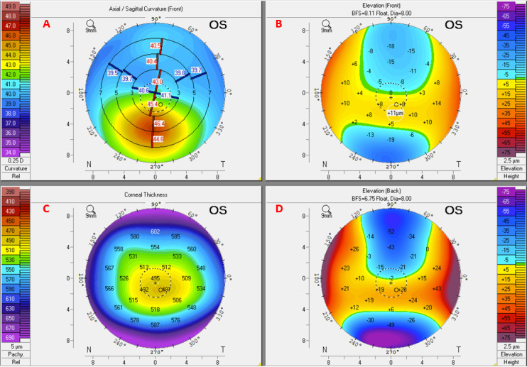
Background: Keratoconus is a disorder characterized by progressive thinning and distortion of the cornea. If detected at an early stage, corneal collagen cross-linking can prevent disease progression and further visual loss. Although advanced forms are easily detected, reliable identification of subclinical disease can be problematic. Several different machine learning algorithms have been used to improve the detection of subclinical keratoconus based on the analysis of multiple types of clinical measures, such as corneal imaging, aberrometry, or biomechanical measurements.
Objective: The aim of this study is to survey and critically evaluate the literature on the algorithmic detection of subclinical keratoconus and equivalent definitions.
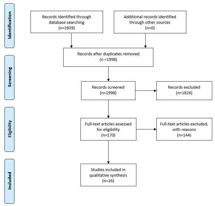
Methods: For this systematic review, we performed a structured search of the following databases: MEDLINE, Embase, and Web of Science and Cochrane Library from January 1, 2010, to October 31, 2020. We included all full-text studies that have used algorithms for the detection of subclinical keratoconus and excluded studies that did not perform validation. This systematic review followed the PRISMA (Preferred Reporting Items for Systematic Reviews and Meta-Analyses) recommendations.
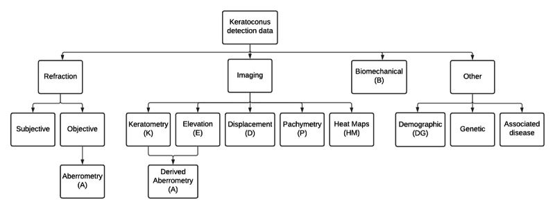
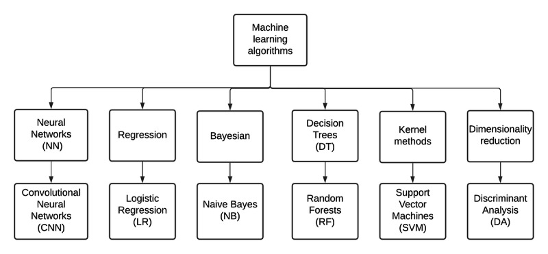
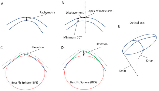
Results: We compared the measured parameters and the design of the machine learning algorithms reported in 26 papers that met the inclusion criteria. All salient information required for detailed comparison, including diagnostic criteria, demographic data, sample size, acquisition system, validation details, parameter inputs, machine learning algorithm, and key results are reported in this study.
Conclusions: Machine learning has the potential to improve the detection of subclinical keratoconus or early keratoconus in routine ophthalmic practice. Currently, there is no consensus regarding the corneal parameters that should be included for assessment and the optimal design for the machine learning algorithm. We have identified avenues for further research to improve early detection and stratification of patients for early treatment to prevent disease progression.
Keywords: artificial intelligence; cornea; corneal disease; corneal imaging; corneal tomography; decision support systems; keratoconus; keratometry; machine learning; subclinical.
©Howard Maile, Ji-Peng Olivia Li, Daniel Gore, Marcello Leucci, Padraig Mulholland, Scott Hau, Anita Szabo, Ismail Moghul, Konstantinos Balaskas, Kaoru Fujinami, Pirro Hysi, Alice Davidson, Petra Liskova, Alison Hardcastle, Stephen Tuft, Nikolas Pontikos. Originally published in JMIR Medical Informatics (https://medinform.jmir.org), 13.12.2021.
Conflict of interest statement
Conflicts of Interest: None declared.
Figures
References
-
- Davidson AE, Hayes S, Hardcastle AJ, Tuft SJ. The pathogenesis of keratoconus. Eye (Lond) 2014 Feb;28(2):189–95. doi: 10.1038/eye.2013.278. http://europepmc.org/abstract/MED/24357835 eye2013278 - DOI - PMC - PubMed
Publication types
Grants and funding
LinkOut - more resources
Full Text Sources
Miscellaneous
